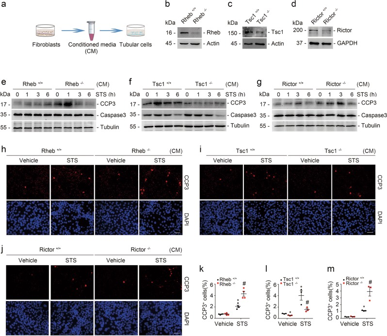
Fig. 5.
Fibroblast mTORC1 and mTORC2 activation protects against staurosporine-induced tubular cell apoptosis. a The coculture system of tubular cells and kidney fibroblasts. b–d Western blot assay showing the abundance for Rheb, Tsc1, and Rictor in kidney fibroblasts. Primary cultured kidney fibroblasts generated from Rhebfl/fl, Tsc1fl/fl, and Rictorfl/fl mice were infected with adeno-Cre for 48 h to induce Rheb, Tsc1, and Rictor gene ablation, respectively. Fibroblasts infected with adeno-GFP were used as control fibroblasts. e–g Western blotting analyses showing the abundance of cleaved caspase 3 in primary cultured tubular cells incubated with CM from various primary cultured kidney fibroblasts as indicated. Tubular cells were treated with staurosporine (1 μM) for different duration. h–j Representative immunofluorescent staining images for cleaved caspase 3 among groups as indicated. Tubular cells were treated with staurosporine (1 μM) for 3 h. Scale bar = 20 µm. k–m Quantitative determination of cleaved caspase 3+ cells among groups as indicated. Data are presented as the percentage of the counted cells. *P < 0.05 versus vehicle-treated Rheb+/+ fibroblasts, n = 4; #P < 0.05 versus staurosporine-treated Rheb+/+ fibroblasts, n = 4. n = 4 refers to four independent repeats (k); *P < 0.05 versus vehicle-treated Tsc1+/+ fibroblasts, n = 3; #P < 0.05 versus staurosporine-treated Tsc1+/+ fibroblasts, n = 3. n = 3 refers to three independent repeats (l); *P < 0.05 versus vehicle-treated Rictor+/+ fibroblasts, n = 3; #P < 0.05 versus staurosporine-treated Rictor+/+ fibroblasts, n = 3. n = 3 refers to three independent repeats (m). CM conditioned media

