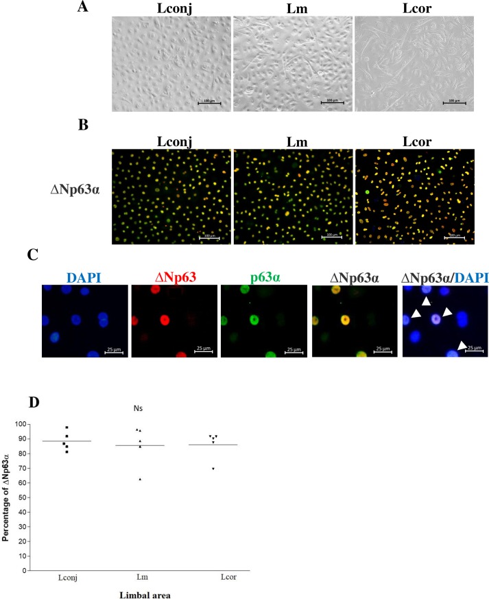
Fig 5. Cell morphology and detection of limbal stem cell marker, ΔNp63α.
(A) Representative confluent cell culture and (B) indirect immunofluorescence detection of the cells expressing both ΔNp63 (red) and p63α (green) that represent ΔNp63α-positive limbal stem cells (yellow) in outgrowth cultures from Lconj, Lm, and Lcor (200× magnification, scale bar = 100 μm). (C) 200× magnification (zoom in) of the stained cells from Lm (scale bar = 25 μm); colocalization of ΔNp63 (red) and p63α (green) was detected in the nuclei (shown in yellow and white arrows). (D) The percent of ΔNp63α-positive cells from Lconj, Lm, and Lcor outgrowth cultures. The line represents the mean percentage of the ΔNp63α-positive cells. Ns, not significant by ANOVA for repeated measures.

