PLOS ONE logoLink to PLOS ONE
. 2020 May 14;15(5):e0224912. doi: 10.1371/journal.pone.0224912

Differences in splicing defects between the grey and white matter in myotonic dystrophy type 1 patients

Masamitsu Nishi 1, Takashi Kimura 1,*, Masataka Igeta 2, Mitsuru Furuta 3, Koichi Suenaga 1,4, Tsuyoshi Matsumura 5, Harutoshi Fujimura 5, Kenji Jinnai 6, Hiroo Yoshikawa 1
Editor: Ruben Artero7
PMCID: PMC7224547  PMID: 32407311

Abstract

Myotonic dystrophy type 1 (DM1) is a multi-system disorder caused by CTG repeats in the myotonic dystrophy protein kinase (DMPK) gene. This leads to the sequestration of splicing factors such as muscleblind-like 1/2 (MBNL1/2) and aberrant splicing in the central nervous system. We investigated the splicing patterns of MBNL1/2 and genes controlled by MBNL2 in several regions of the brain and between the grey matter (GM) and white matter (WM) in DM1 patients using RT-PCR. Compared with amyotrophic lateral sclerosis (ALS, as disease controls), the percentage of spliced-in parameter (PSI) for most of the examined exons were significantly altered in most of the brain regions of DM1 patients, except for the cerebellum. The splicing of many genes was differently regulated between the GM and WM in both DM1 and ALS. In 7 out of the 15 examined splicing events, the level of PSI change between DM1 and ALS was significantly higher in the GM than in the WM. The differences in alternative splicing between the GM and WM may be related to the effect of DM1 on the WM of the brain.

Introduction

Myotonic dystrophy type 1 (DM1) is the most common form of muscular dystrophy in adults, affecting the skeletal muscle, heart, ocular lens, testis, and central nervous system (CNS). The CNS symptoms of DM1 can have a negative impact on a patient’s quality of life [1]. DM1 is caused by the unstable expansion of CTG trinucleotide repeats in the 3’ untranslated region (UTR) of the myotonic dystrophy protein kinase (DMPK) gene. These CTG repeats are transcribed to CUG repeats, leading to the formation of an RNA hairpin loop. These loops form foci in the nucleus and sequester splicing factors, such as the muscleblind-like (MBNL) proteins. The MBNL proteins control splicing; in the nucleus, MBNL binds to the YGCY motif of pre-mRNAs and controls the splicing of alternative exons in either an exon inclusion or exon exclusion direction, depending on whether the motif is located in the downstream or upstream intron for each exon, respectively [2]. We have previously detected several splicing defects in the brains of both MBNL1/2 knockout mice and DM1 patients, and revealed that MBNL2 is a major splicing factor in the brain, while MBNL1 performs a similar function in the skeletal muscle [3, 4]. In addition, conditional Mbnl1 and Mbnl2 double-knockout mice showed greater splicing defects than either Mbnl1 or Mbnl2 knockout, indicating that the combined loss of MBNL1 and MBNL2 is necessary to exacerbate mis-splicing, since MBNL1 and MBNL2 compensate for each other [5, 6].

We have previously examined the mis-splicing in DM1 patients for each area of the brain and found that mis-splicing in the cerebellum is less apparent than the other areas [3]. We also demonstrated the different degrees of mis-splicing that occurs among cell layers of the cerebellum [7]. These results clearly showed the heterogeneity of the brain and show that detailed splicing analysis for each cell or region is essential to clarify the pathomechanisms of this disorder.

It has been reported that fetal splice isoforms increase in adult DM1 tissues as a result of MBNL1/2 sequestration, which regulates the splicing switch from fetal to adult [4, 811]. MBNL2 splicing is developmentally regulated; exon 5 (54nt.) and exon 8 (95nt.) are included in the fetal brain, while both exons are excluded in the adult brain [12].

Using RNA-sequencing, Mills et al. showed that the grey matter (GM) and white matter (WM) have distinct transcriptome profiles, including alternative splicing (AS) [13]. Neuroimaging analysis in patients with DM1 revealed various changes with conventional MRI [14, 15], voxel-based morphometry (VBM) [16, 17], and diffusion-weighted imaging (DWI) tensor analysis [1821]. Overall, the degree of change is greater in the WM than in the GM [17, 22].

Aberrant splicing of MBNL1/2 in the brain of DM1 patients and mouse models has been previously reported [2325]. Considering distinct regions are affected, as shown by neuroimaging, and the differences in splicing regulation between the GM and WM, it is important to determine how AS is controlled in GM and WM of a DM1 brain. In this study, we examined the splicing patterns of MBNL1/2, and the other genes controlled by MBNL2, among several brain regions (frontal and temporal lobes, hippocampus, the cerebellum), and between the GM and WM in DM1 patients.

Materials and methods

Ethics and written informed consent

This research was approved by the Ethics Committee of Hyogo College of Medicine (approval number: 93), and written informed consent was obtained from patients themselves or from their family members for an autopsy.

MBNL1/2 and APP DNA sequence and primer design

We searched the DNA sequences for MBNL1, MBNL2 and APP using the GENETYX® NCBI database. MBNL1 has over 50 variants depending on the presence or absence of exons 5 (54 nt.), 6 (154 nt.), 7 (36 nt.), 8 (95 nt.), and 9 (64 nt.), or changes in the non-coding region. MBNL2 has 10 splicing variants depending on the presence or absence of exons 5 (54 nt.), 7 (36 nt.), and 8 (95 nt.). APP has 3 variants depending on the presence or absence of exon 7 (168 nt.) and exon 8 (57 nt.). Using NCBI Primer-BLAST, we designed two sets of primers for each spliced gene; MBNL1/2 exon 4 forward and exon 6 reverse primers for exon 5 splicing, and exon 6 forward and exon 9 reverse primers for exons 7 and 8 splicing. APP exon 6 forward and exon 9 reverse primers for exon 7 and 8 splicing. In addition, we examined splicing patterns of other genes controlled by MBNL2: ADD1 exon 15 (34 nt.), CACNA1D exon 11 (60 nt.), CLASP2 exon 23a (27 nt.) or 23b (27nt.), CSCNK1D exon 9 (64 nt.), GRIN1 exon 4 (63 nt.), KCNMA1 exon 27a (81 nt.), MAPT exon 2 (87 nt.) and exon 10 (93 nt.), TANC2 exon 22a (30 nt.), using the primers used in our previous report [3, 7] (Table 1).

Table 1. RT-PCR primers for alternative splicing.

Genes (target exon) Forward primer Reverse primer
ADD1 (exon 15 (34 nt.)) GGACGAGGCTAGAGAACAGAAAGAAAAGA TTGGGAAGCCGAGTGCTTCTGAA
APP (exon 7 (168 nt.), exon 8 (57 nt.)) TCTGTGGAAGAGGTGGTTCG TGGCCTCAAGCCTCTCTTTG
CACNA1D (exon 12 (60 nt.)) CACAGAGAACGTCAGCGGT TGAGTTTGGATTTTGAGATGGC
CLASP2 (exon 23a (27 nt.), exon 23b (27 nt.)) GCTGGCATGGGAAATGCCAAGGC GCTCCGTGGTATCTTGCTTCTTTTT
CSNK1D (exon 9 (64 nt.)) GATACCTCTCGCATGTCCACCTCACA GCATTGTCTGCCCTTCACAGCAAT
GRIN1 (exon 4 (63 nt.)) GTCTACAGCTGGAACCACATC TCCATCAGCAGGGCCGTCACG
KCNMA1 (exon 27a (81 nt.)) CGTTCACACCTCCAGGAATGGATAGAT GTGAGGTACAGTTCTGTATCAGGGTCAT
MAPT (exon 2 (87 nt.)) TACACCATGCACCAAGACCA GTCTCCAATGCCTGCTTCTT
MAPT (exon 10 (93 nt.)) ACTGAGAACCTGAAGCACCAG CACTTGGAGGTCACCTTGCTC
MBNL1 (exon 5 (54 nt.)) TCAAGGCTGCCCAATACCAG TGTTGGCTAGAGCCTGTTGG
MBNL1 (exon 7 (36 nt.), exon 8 (95 nt.)) ACCAACAGGCTCTAGCCAAC GGCTAGTCAGATGTTCGGCA
MBNL2 (exon 5 (54 nt.)) AGGCCAAAATCAAAGCTGCG GTGAGAGCCTGCTGGTAGTG
MBNL2 (exon 7 (36 nt.), exon 8 (95 nt.)) CACGCCGCGTTCATTCCAAC TAGCATGCAGTTTGTGGCAA
TANC2 (exon 22a (30 nt.)) GCCATGATCGAGCACGTTGACTACAGT CCTCTTCCATCAGCTTGCTCAACA

Human RNA and splicing analysis

We investigated brains of patients during autopsy, with RNA extracted from the brain of 6 patients with DM1 and 6 patients with amyotrophic lateral sclerosis (ALS, as disease controls). The clinical features of samples are summarized in Table 2 [26]. Two RNA samples of fetal brains (as fetal controls) were obtained commercially (Cat. No. 540157, Agilent Technologies, US; Cat. No. 636526, Takara Bio Inc. Japan). RNA was extracted from the frontal and temporal lobes, the hippocampus, and the cerebellum using the ISOGEN® reagent (Nippon Gene, Japan). The splicing patterns in the different brain regions and between the GM and WM.

Table 2. Clinical features of samples.

Disease Gender Age Onset age Duration Type of DM1 Cognitive decline
DM1 F 58 38 20 years Adult onset Mild
F 66 42 24 years Late onset Mild
M 47 12 35 years Juvenile onset Severe
M 65 40 25 years Adult onset Mild
M 78 41 37 years Late onset Mild
M 57 18 39 years Juvenile onset Mild
ALS F 53 N/A N/A N/A Normal
M 69 N/A N/A N/A Normal
M 67 N/A N/A N/A Normal
F 73 N/A N/A N/A Normal
M 70 N/A N/A N/A Normal

Type of DM1; Juvenile onset (age at onset 10–20 years), Adult onset (age at onset 20–40 years), Late onset (age at onset > 40 years). The degree of cognitive decline was assessed on a three-point scale; Normal, Mild, and Severe.

For GM and WM, the frontal lobe tissue was sliced into 20-μm thick sections using a Cryostat (Leica® CM1520, Germany). Before separation, one of the sections was stained using the Luxol Fast Blue Stain Kit (ScyTek Laboratories Inc., US), to identify the boundary between the GM and WM. Only unstained sections were used for RNA extraction. We manually separated each section into the GM and WM and extracted RNA using RNeasy® Plus Mini (QIAGEN®, Germany) for comparison between the GM and WM.

To ensure enrichment of the GM and WM, we used the primers for NEFH (higher expression in the GM than the WM), MOG (higher expression in the WM than the GM), and GAPDH (control), and examined this by quantitative real-time PCR (qPCR) using the PowerUp SYBR with the 7500 Real-Time PCR system (Applied Biosystems, US) (Table 3). qPCR was performed in triplicates [13]. The levels of NEFH and MOG mRNA expression were calculated by the 2-ΔΔCt method, using GAPDH as an endogenous control, and the data were presented as the mean of triplicates.

Table 3. Quantitative RT-PCR primers for confirming the separation between the GM and WM.

Genes Forward primer Reverse primer
NEFH AGGTGAAGAGTGTCGGATTG GAAGCGAGAAAGGAATTGGG
MOG CCTCCACTTGGCCTGACCTT ACCTCCATGCCTGTAGCGTT
GAPDH CCATCACTGCCACCCAGAAGAC CCATCACGCCACAGTTTCCC

All cDNA was synthesized using the extracted RNA and purchased RNA (1 μg of RNA was used for comparison among several brain regions; 10–400 ng of RNA was used for comparison between the GM and WM). Random hexamers with the SuperScript® III First-Strand Synthesis System (Invitrogen, US) for reverse transcription PCR (RT-PCR) were used according to manufacturer’s instructions (Applied Biosystems). cDNA was amplified using AmpliTaq Gold® 360 Master Mix (Applied Biosystems), with the initial denaturation set at 94°C for 10 min, followed by 32 (for comparison among several brain regions) or 36 (for comparison between the GM and WM) amplification cycles at 94°C for 30 s, 61°C for 30 s, and 72°C for 60 s. PCR products were analyzed with an Agilent 2100 Bioanalyzer (Agilent Technologies, US). The linearity of the PCR amplification at each cycle was verified with the DM1 and ALS samples. We used percent spliced-in (PSI) values, indicating inclusion ratio of an alternative exon [3].

Statistical analysis

The Welch’s t-test was performed for testing mean difference of PSI between ALS and DM1 with two-sided significant level of 5% for all comparisons. No adjustment of significant level to multiplicity was applied to retain maximum power in this study, as suggested by Saville [27]. Since some data showed skew distribution, we evaluated the impact of the assumption of the normality of the Welch’s t-test via sensitivity analyses as following steps; First, we performed the Shapiro-Wilk test for testing the normality. Then if the test of normality was significant at least one group within a group comparison, the Wilcoxon rank sum test was applied to the group comparison. Only the p-values of the Welch’s t-test were shown in Figures since we confirmed the consistent significant results between the Welch’s t-test and the Wilcoxon rank sum test for the cases where the test of normality was significant (data not shown). We calculated the average level of PSI change (ΔPSI), which was obtained by subtracting the average of PSI of the ALS from the DM1. The summary statistics, 95% confidence interval based on the unpooled variance, and p-value calculated using Welch’s t-test for the difference between average ΔPSI GM and average ΔPSI WM for each exon are provided. The confidence interval and p-value were evaluated for the difference between average PSI DM1 (GM-WM) and average PSI control (GM-WM) because these differences were mathematically equivalent in our data, which had no missing within-subject GM and WM PSIs for each exon.

Results

Comparison among several brain regions

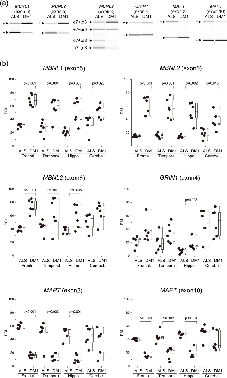
Compared with the ALS, PSI for MBNL1 exon 5 were higher in all examined brain areas of DM1. The PSI for MBNL1 exon 8 were significantly higher in the DM1 temporal lobe than in the ALS (Fig 1, S1 Fig). The result of MBNL1 exon 5 splicing was similar to a previous report [23], and that of exon 8 was not reported. Compared with the ALS, PSI for MBNL2 exon 5 were higher in all examined brain areas of DM1 and exon 8 were significantly higher in most brain areas of DM1, except for the cerebellum. Notably, for the frontal and temporal regions, and hippocampus of DM1, PSI for MBNL1 and MBNL2 exons 5 and exon 8 were highly variable compared with the ALS.

Fig 1. Aberrant splicing among several regions of the brain.

Fig 1

(a) Representative RT-PCR products from the frontal lobe in the ALS and DM1. (b) Inclusion ratios of splicing changes in several brain regions. PSI values of all examined genes were compared by Welch’s T-test. In the box-and-whisker plot, the line inside the box represents the median and the square symbol represents the average. ALS, amyotrophic lateral sclerosis; DM1, myotonic dystrophy type 1; Frontal., Frontal lobe; Temporal., Temporal lobe; Hippo., Hippocampus; Cerebel., Cerebellum.

Compared with the ALS, PSI for MAPT exon 2 and exon 10 were significantly lower in most brain areas of DM1, except for the cerebellum. PSI for GRIN1 exon 4 was significantly higher in the DM1 hippocampus than in the ALS. Other areas had no significant difference. PSI was very low in the fetal brain tissue (Fig 1, S2 Fig).

Comparison between the GM and WM

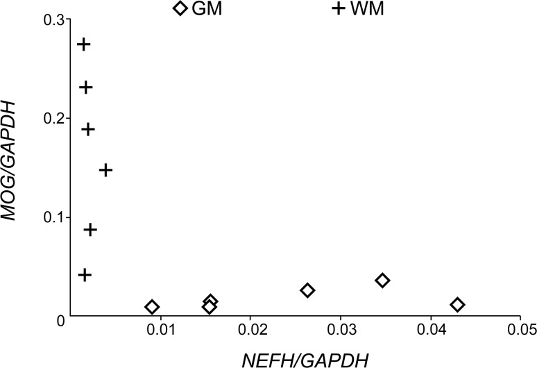
Q-PCR analysis showed that the expression levels of NEFH was higher in the GM, while that of MOG was higher in the WM (Fig 2), confirming that samples were enriched for the GM and WM.

Fig 2. Expression level of NEFH and MOG in the GM and WM.

Fig 2

Concentration of NEFH and MOG mRNA calculated by the 2-ΔΔCt method, which was divided by that of GAPDH. Data are presented as the mean of triplicates. GM, grey matter; WM, white matter.

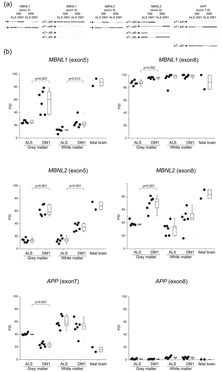
There were three patterns of AS in the fetal brain: 1) exon inclusion type (PSI ≥ 60%, ADD1, CLASP2, MBNL1/2), 2) exon exclusion type (PSI < 40%, APP, CACNA1D, CSNK1D, GRIN1, KCNMA1, MAPT) (Fig 3, S2 Fig), and 3) moderate type (40% ≤ PSI < 60%, TANC2).

Fig 3. Aberrant splicing between the GM and WM.

Fig 3

(a) Representative RT-PCR products from the frontal lobe GM and WM in ALS and DM1. (b) Inclusion ratios of splicing changes in the GM and WM. PSI values of all examined genes were compared by Welch’s T-test. In the box-and-whisker plot, the line inside the box represents the median and the square symbol represents the average. ALS, amyotrophic lateral sclerosis; DM1, myotonic dystrophy type 1; GM, grey matter; WM, white matter.

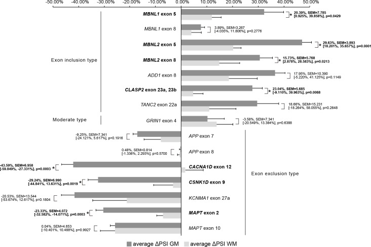
We used ΔPSI to compare splicing defects between GM and WM. For example, average PSI of MBNL1 for exon 5 were 57.43%, 24.33%, 25.06%, and 12.35% in the GM and WM of DM1, and the GM and WM of ALS, respectively. ΔPSI of the GM was 32.38%, while ΔPSI of the WM was 11.99%. In 7 (MBNL1 exon 5, MBNL2 exon 5, MBNL2 exon 8, CLASP2 exon 23a or 23b, CACNA1D exon 12, CSNK1D exon 9, MAPT exon 2), out of 15 examined splicing events, | ΔPSI | of the GM was significantly higher than that of the WM, suggesting that more splicing misregulation occurs in the GM (Fig 4).

Fig 4. Comparison of ΔPSI GM and ΔPSI WM.

Fig 4

This bar graph shows average ΔPSI GM and average ΔPSI WM. The error bars represent SEM. Average Δ PSI values of all examined genes were compared by Welch’s T-test. The numbers in the graph indicate the mean difference, SEM, [Lower CL, Upper CL], and p-value in this order for comparison between average ΔPSI GM and average ΔPSI WM. The gene names and numbers with statistical significance were written in bold with asterisk. GM, grey matter; WM, white matter.

PSI of APP exon 8 and GRIN1 exon 4 showed no statistically significant difference between DM1 and the ALS (Fig 3, S2 Fig).

Discussion

Comparisons among the brain regions revealed that PSI of almost all examined gene exons were significantly different in the DM1 patients compared with the ALS, except for the cerebellum. Comparison between the GM and WM revealed that the splicing of many genes was differently regulated between the GM and WM. The extent of splicing change between the DM1 and ALS was higher in the GM than in the WM.

According to our previous study, there were fewer alternative splicing defects in the DM1 cerebellum than in other brain regions [3]. We showed that the inclusion of MBNL1 exon 5 and MBNL2 exon 5 and exon 8 was higher in most brain areas, except the cerebellum for MBNL2 exon 5 and exon 8. MBNL1/2 exon 5 inclusion in the DM1 human brain had previously been reported [23, 24]; however, we are the first to demonstrate MBNL2 exon 8 inclusion in the DM1 human brain. MBNL1/2 splicing in several brain regions were similar to previous reports [3], in that aberrant splicing is observed in most brain areas except for the cerebellum.

MAPT [28] and GRIN1 [10] splicing defects were found in DM1 patients’ brains. The aberrant splicing of MAPT exons 2, 3 [4], 10 and GRIN1 [5] were recapitulated by using knock out mice of Mbnl1/2, suggesting that these splicing events are controlled by the MBNL 1/2 protein. We analyzed both these genes in several areas of the brain and found that MAPT splicing was similar to that of other genes, where there were fewer splicing changes in the cerebellum than in other brain areas in the DM1. GRIN1 was different in that: 1) In the hippocampus, PSI of the DM1 was significantly higher than that of ALS. In the frontal and temporal lobes, PSI of the DM1 tended to be higher than that of the ALS, but these differences did not reach statistical significance; 2) PSI in the fetal brain was lower than that in the ALS, in contrast to the DM1. A significant difference was observed in the hippocampus between the ALS and DM1, but not in other areas. Interestingly, the splicing defect of GRIN1 in the hippocampus does not represent a return to an embryonic splicing profile. However, ΔPSI is very low (~ 5%), and thus the functional consequences of such a minor difference might be minimal. It is difficult to draw a conclusion without further examination.

Analysis between the GM and WM revealed that, compared to the ALS, splicing changes in the GM of DM1 patients exceeded those in WM, in 7 out of the 15 examined exons. When several brain regions were compared, we collected RNA from both the GM and WM, and did not evaluate the ratio of GM to WM. Therefore, we assume that the distribution of PSI observed in the comparison among several brain regions may have been caused by the variation in the ratio of GM/WM in the collected samples.

This study revealed that the degree of mis-splicing in DM1 differs between the GM and WM. The GM is composed of the neuronal cell bodies, protoplasmic astrocytes, and microglial cells, while the WM comprises axons, oligodendrocytes, fibrous astrocytes, microglial cells, and ependymal cells (near the brain ventricle) [29]. In this study, as mRNA was extracted from the WM just under the GM, ependymal cells were not included in the samples. There are a few possibilities that could explain why these differences in splicing between the GM and WM occurred. One possibility is that these differences may be affected by the distribution of each splice isoform between the axon and neuronal cell body. The WM is less transcriptionally active than the GM, and mRNA in the WM may represent mRNA flow from the GM [13], since the mRNAs are transcribed in the neuronal cell body, and part of them are transported to the axon [30]. MBNL1/2 exon 5 includes nuclear localization signals, and it is reported that the isoforms with this exon tend to be exclusively expressed in the nucleus [12]. Inclusion of exon 8 reduces the hairpin loop mobility of MBNL2 [12]. Since both exons tend to be present in DM1 patients, there is less MBNL in the cytoplasm. In order to target to specific compartments, MBNL binds to the distal 3’UTR protein binding sites and may intercede isoform specific mRNA localization [31]. Hence, it is possible that the presence or absence of each alternative exon may influence its binding ability to the 3’UTR of MBNL and the transport of its mRNA. Consequently, aberrant splicing of MBNL mRNA and the resulting intracellular localization shift of MBNL in DM1 patients might affect axonal transport of each spliced mRNA isoform. Another possibility is that splicing differences between the GM and WM may be influenced by splicing patterns of other cells such as oligodendrocytes, fibrous astrocytes, and microglial cells of the WM. Jiang showed that in DM1 patients, PSI for APP exon 7 were lower than in the control [10]. APP exon 7 and 8 are excluded from the neuronal APP isoform (APP 695), but are included in the astrocytic APP isoform (APP 751, 770) [32]. We showed that in the GM, PSI for APP exon 7 in the DM1 was significantly lower than in the ALS, but there was no statistical significance in WM. From this result, we presumed that the effect of aberrant splicing of astrocyte in the WM may be negligible. These distinct splicing changes may be due to differences in the number of CTG repeats in the cells between the GM and WM. Jinnai et al. examined the somatic instability of the CTG expansion in various regions of the CNS and found that the expansion in the GM was longer than that in the WM, although this difference did not reach statistical significance [33]. Possibly, this relatively short expansion in the WM might explain the modest level of aberrant splicing. In order to examine this possibility, it is necessary to isolate each cell type using cell culture or laser microdissection.

VBM and DWI tensor studies have shown that the effect of DM1 on the WM is more prominent than on the GM and that WM T2 hyperintensity lesions were found in the frontal, temporal (especially at the anterior temporal poles), and parietal lobes [21, 22]. However, this study showed more splicing defects in the GM than in the WM. It is unclear how these differences in the degree of splicing abnormalities relate to the predominant WM change. It remains unclear if the change in the WM is a result of Wallerian degeneration or a primary process of DM1 [17, 19]. As mentioned above, we suggested two possibilities explaining the differences in splicing defects between the GM and WM. Assuming the possibility that aberrant/fetal splicing mRNA isoforms are difficult to transport to the axon, aberrantly spliced isoforms would increase in the neuronal cell body, which could cause axonal injury as a consequence of Wallerian degeneration. The possibility that fewer splicing defects occurred in the myelin sheath of oligodendrocytes compared to the neuronal cell body does not explain the predominantly affected WM as shown by neuroimaging. Taken together, we hypothesize that WM lesions were caused by Wallerian degeneration due to neuronal cell body damage. Although there are limited findings on how each splice isoform protein is distributed, this is the first study showing the differences in splicing regulation and the degree of aberrant splicing between the GM and WM in DM1 patients.

Limitations

This study had some limitations. First, we used ALS brain samples as the disease control. ALS tissues show splicing deregulation [34] and ALS brains may also display significant changes in WM and GM [35]. Our previous study showed statistical significance in the splicing defects in the temporal lobe of DM1 compared with either disease controls (7 out of 9 samples were from ALS) or healthy controls [4]. However, other areas, GM and WM of healthy controls have not been examined, and further study is needed in the future. Second, although it is important to determine whether the degree of mis-splicing, as the difference between the GM and WM, is related to each type of DM1 and the degree of cognitive decline, we could not determine a clear trend in this study, probably because the sample size was small and the variability of the PSI values between samples was large.

Conclusion

In this study, we showed that splicing changes are relatively modest in the WM compared to the GM in the brain of DM1, which indicated the possibility that aberrant/fetal splicing isoforms may not be transported to the axon. Our result suggests that the predominant effects of DM1 on the WM might represent axonal injury by Wallerian degeneration due to GM dominant aberrant splicing. We believe that exploring the distribution of each spliced protein isoform by immunostaining can help in our understanding of the pathological mechanisms of DM1. Future studies should also investigate mRNA and protein transport and local translation in the axons of neuronal cells.

Supporting information

S1 Fig. MBNL1 exon 8 aberrant splicing among several regions of the brain.

Inclusion ratios of splicing changes in several brain regions. PSI values of MBNL1 exon 8 was compared by Welch’s T-test. In box-and-whisker plot, the line inside the box is the median, square symbol is the average. ALS, amyotrophic lateral sclerosis; DM1, myotonic dystrophy type 1; Frontal., Frontal lobe; Temporal., Temporal lobe; Hippo., Hippocampus; Cerebel., Cerebellum.

(TIF)

S2 Fig. Aberrant splicing between the GM and WM.

Inclusion ratios of splicing changes in the GM and WM. PSI values of all examined genes were compared by Welch’s T-test. In box-and-whisker plot, the line inside the box is the median, square symbol is the average. ALS, amyotrophic lateral sclerosis; DM1, myotonic dystrophy type 1.

(TIF)

Acknowledgments

The authors thank the Research Resource network Japan for the human brain samples and editage (www.editage.jp) for English language editing.

Data Availability

All relevant data are within the manuscript and its Supporting Information files.

Funding Statement

This work was supported by JSPS KAKENHI Grant Number JP18K07515. The funders had no role in study design, data collection and analysis, decision to publish, or preparation of the manuscript.

References

  • 1.Bugiardini E, Meola G, Group D-C. Consensus on cerebral in-volvement in myotonic dystrophy: workshop report: May 24–27, 2013, Ferrere (AT), Italy. Neuromuscul Disord. 2014;24(5):445–52. 10.1016/j.nmd.2014.01.013 [DOI] [PubMed] [Google Scholar]
  • 2.Zhang C, Lee KY, Swanson MS, Darnell RB. Prediction of clus-tered RNA-binding protein motif sites in the mammalian genome. Nucleic Acids Res. 2013;41(14):6793–807. 10.1093/nar/gkt421 [DOI] [PMC free article] [PubMed] [Google Scholar]
  • 3.Suenaga K, Lee KY, Nakamori M, Tatsumi Y, Takahashi MP, Fu-jimura H, et al. Muscleblind-like 1 knockout mice reveal novel splicing defects in the myotonic dystrophy brain. PLoS One. 2012;7(3):e33218 10.1371/journal.pone.0033218 [DOI] [PMC free article] [PubMed] [Google Scholar]
  • 4.Charizanis K, Lee KY, Batra R, Goodwin M, Zhang C, Yuan Y, et al. Muscleblind-like 2-mediated alternative splicing in the developing brain and dysregulation in myotonic dystrophy. Neuron. 2012;75(3):437–50. 10.1016/j.neuron.2012.05.029 [DOI] [PMC free article] [PubMed] [Google Scholar]
  • 5.Goodwin M, Mohan A, Batra R, Lee KY, Charizanis K, Fernández Gómez FJ, et al. MBNL Sequestration by Toxic RNAs and RNA Mispro-cessing in the Myotonic Dystrophy Brain. Cell Rep. 2015;12(7):1159–68. 10.1016/j.celrep.2015.07.029 [DOI] [PMC free article] [PubMed] [Google Scholar]
  • 6.Lee KY, Li M, Manchanda M, Batra R, Charizanis K, Mohan A, et al. Compound loss of muscleblind-like function in myotonic dystrophy. EMBO Mol Med. 2013;5(12):1887–900. 10.1002/emmm.201303275 [DOI] [PMC free article] [PubMed] [Google Scholar]
  • 7.Furuta M, Kimura T, Nakamori M, Matsumura T, Fujimura H, Jinnai K, et al. Macroscopic and microscopic diversity of missplicing in the central nervous system of patients with myotonic dystrophy type 1. Neuroreport. 2018;29(3):235–40. 10.1097/WNR.0000000000000968 [DOI] [PubMed] [Google Scholar]
  • 8.Braz SO, Acquaire J, Gourdon G, Gomes-Pereira M. Advances in the Understanding of Neuromuscular Aspects of Myotonic Dystrophy. Front Neurol. 2018;9:519 10.3389/fneur.2018.00519 [DOI] [PMC free article] [PubMed] [Google Scholar]
  • 9.Lin X, Miller JW, Mankodi A, Kanadia RN, Yuan Y, Moxley RT, et al. Failure of MBNL1-dependent post-natal splicing transitions in myo-tonic dystrophy. Hum Mol Genet. 2006;15(13):2087–97. 10.1093/hmg/ddl132 [DOI] [PubMed] [Google Scholar]
  • 10.Jiang H, Mankodi A, Swanson MS, Moxley RT, Thornton CA. Myotonic dystrophy type 1 is associated with nuclear foci of mutant RNA, sequestration of muscleblind proteins and deregulated alternative splicing in neurons. Hum Mol Genet. 2004;13(24):3079–88. 10.1093/hmg/ddh327 [DOI] [PubMed] [Google Scholar]
  • 11.Kanadia RN, Johnstone KA, Mankodi A, Lungu C, Thornton CA, Esson D, et al. A muscleblind knockout model for myotonic dystrophy. Science. 2003;302(5652):1978–80. 10.1126/science.1088583 [DOI] [PubMed] [Google Scholar]
  • 12.Sznajder Ł, Michalak M, Taylor K, Cywoniuk P, Kabza M, Wojtkowiak-Szlachcic A, et al. Mechanistic determinants of MBNL activi-ty. Nucleic Acids Res. 2016;44(21):10326–42. 10.1093/nar/gkw915 [DOI] [PMC free article] [PubMed] [Google Scholar]
  • 13.Mills JD, Kavanagh T, Kim WS, Chen BJ, Kawahara Y, Halliday GM, et al. Unique transcriptome patterns of the white and grey matter cor-roborate structural and functional heterogeneity in the human frontal lobe. PLoS One. 2013;8(10):e78480 10.1371/journal.pone.0078480 [DOI] [PMC free article] [PubMed] [Google Scholar]
  • 14.Abe K, Fujimura H, Toyooka K, Yorifuji S, Nishikawa Y, Hazama T, et al. Involvement of the central nervous system in myotonic dystrophy. J Neurol Sci. 1994;127(2):179–85. 10.1016/0022-510x(94)90071-x [DOI] [PubMed] [Google Scholar]
  • 15.Ogata A, Terae S, Fujita M, Tashiro K. Anterior temporal white matter lesions in myotonic dystrophy with intellectual impairment: an MRI and neuropathological study. Neuroradiology. 1998;40(7):411–5. 10.1007/s002340050613 [DOI] [PubMed] [Google Scholar]
  • 16.Schneider-Gold C, Bellenberg B, Prehn C, Krogias C, Schneider R, Klein J, et al. Cortical and Subcortical Grey and White Matter Atrophy in Myotonic Dystrophies Type 1 and 2 Is Associated with Cognitive Im-pairment, Depression and Daytime Sleepiness. PLoS One. 2015;10(6):e0130352 10.1371/journal.pone.0130352 [DOI] [PMC free article] [PubMed] [Google Scholar]
  • 17.Minnerop M, Weber B, Schoene-Bake JC, Roeske S, Mirbach S, Anspach C, et al. The brain in myotonic dystrophy 1 and 2: evidence for a predominant white matter disease. Brain. 2011;134(Pt 12):3530–46. 10.1093/brain/awr299 [DOI] [PMC free article] [PubMed] [Google Scholar]
  • 18.Fukuda H, Horiguchi J, Ono C, Ohshita T, Takaba J, Ito K. Diffu-sion tensor imaging of cerebral white matter in patients with myotonic dystrophy. Acta Radiol. 2005;46(1):104–9. 10.1080/02841850510015974 [DOI] [PubMed] [Google Scholar]
  • 19.Ota M, Sato N, Ohya Y, Aoki Y, Mizukami K, Mori T, et al. Rela-tionship between diffusion tensor imaging and brain morphology in pa-tients with myotonic dystrophy. Neurosci Lett. 2006;407(3):234–9. 10.1016/j.neulet.2006.08.077 [DOI] [PubMed] [Google Scholar]
  • 20.Wozniak JR, Mueller BA, Ward EE, Lim KO, Day JW. White matter abnormalities and neurocognitive correlates in children and adoles-cents with myotonic dystrophy type 1: a diffusion tensor imaging study. Neuromuscul Disord. 2011;21(2):89–96. 10.1016/j.nmd.2010.11.013 [DOI] [PMC free article] [PubMed] [Google Scholar]
  • 21.Okkersen K, Monckton DG, Le N, Tuladhar AM, Raaphorst J, van Engelen BGM. Brain imaging in myotonic dystrophy type 1: A systematic review. Neurology. 2017;89(9):960–9. 10.1212/WNL.0000000000004300 [DOI] [PubMed] [Google Scholar]
  • 22.Minnerop M, Gliem C, Kornblum C. Current Progress in CNS Imaging of Myotonic Dystrophy. Front Neurol. 2018;9:646 10.3389/fneur.2018.00646 [DOI] [PMC free article] [PubMed] [Google Scholar]
  • 23.Dhaenens CM, Schraen-Maschke S, Tran H, Vingtdeux V, Ghanem D, Leroy O, et al. Overexpression of MBNL1 fetal isoforms and modified splicing of Tau in the DM1 brain: two individual consequences of CUG trinucleotide repeats. Exp Neurol. 2008;210(2):467–78. 10.1016/j.expneurol.2007.11.020 [DOI] [PubMed] [Google Scholar]
  • 24.Hernández-Hernández O, Sicot G, Dinca DM, Huguet A, Nicole A, Buée L, et al. Synaptic protein dysregulation in myotonic dystrophy type 1: Disease neuropathogenesis beyond missplicing. Rare Dis. 2013;1:e25553 10.4161/rdis.25553 [DOI] [PMC free article] [PubMed] [Google Scholar]
  • 25.Sicot G, Servais L, Dinca DM, Leroy A, Prigogine C, Medja F, et al. Downregulation of the Glial GLT1 Glutamate Transporter and Purkinje Cell Dysfunction in a Mouse Model of Myotonic Dystrophy. Cell Rep. 2017;19(13):2718–29. 10.1016/j.celrep.2017.06.006 [DOI] [PMC free article] [PubMed] [Google Scholar]
  • 26.Gourdon G, Meola G. Myotonic Dystrophies: State of the Art of New Therapeutic Developments for the CNS. Front Cell Neurosci. 2017;11:101 10.3389/fncel.2017.00101 [DOI] [PMC free article] [PubMed] [Google Scholar]
  • 27.Saville DJ. Multiple Comparison Procedures: The Practical Solu-tion. The American Statistician. 1990;44(2):174–80. [Google Scholar]
  • 28.Sergeant N, Sablonnière B, Schraen-Maschke S, Ghestem A, Maurage CA, Wattez A, et al. Dysregulation of human brain microtubule-associated tau mRNA maturation in myotonic dystrophy type 1. Hum Mol Genet. 2001;10(19):2143–55. 10.1093/hmg/10.19.2143 [DOI] [PubMed] [Google Scholar]
  • 29.Mescher AL, Junqueira LCU. Junqueira's basic histology: texts and atlas. Fifteenth edition ed. New York: McGraw Hill Education; 2018. ix, 562 pages p. [Google Scholar]
  • 30.Cioni JM, Koppers M, Holt CE. Molecular control of local trans-lation in axon development and maintenance. Curr Opin Neurobiol. 2018;51:86–94. 10.1016/j.conb.2018.02.025 [DOI] [PubMed] [Google Scholar]
  • 31.Wang ET, Cody NA, Jog S, Biancolella M, Wang TT, Treacy DJ, et al. Transcriptome-wide regulation of pre-mRNA splicing and mRNA lo-calization by muscleblind proteins. Cell. 2012;150(4):710–24. 10.1016/j.cell.2012.06.041 [DOI] [PMC free article] [PubMed] [Google Scholar]
  • 32.Caillet-Boudin ML, Fernandez-Gomez FJ, Tran H, Dhaenens CM, Buee L, Sergeant N. Brain pathology in myotonic dystrophy: when tauopathy meets spliceopathy and RNAopathy. Front Mol Neurosci. 2014;6:57 10.3389/fnmol.2013.00057 [DOI] [PMC free article] [PubMed] [Google Scholar]
  • 33.Jinnai K, Mitani M, Futamura N, Kawamoto K, Funakawa I, Itoh K. Somatic instability of CTG repeats in the cerebellum of myotonic dys-trophy type 1. Muscle Nerve. 2013;48(1):105–8. 10.1002/mus.23717 [DOI] [PubMed] [Google Scholar]
  • 34.Butti Z, Patten SA. RNA Dysregulation in Amyotrophic Lateral Sclerosis. Front Genet. 2018;9:712 10.3389/fgene.2018.00712 [DOI] [PMC free article] [PubMed] [Google Scholar]
  • 35.Zhou T, Ahmad TK, Gozda K, Truong J, Kong J, Namaka M. Implications of white matter damage in amyotrophic lateral sclerosis (Review). Mol Med Rep. 2017;16(4):4379–92. 10.3892/mmr.2017.7186 [DOI] [PMC free article] [PubMed] [Google Scholar]

Decision Letter 0

Ruben Artero

16 Nov 2019

PONE-D-19-29543

Differences in splicing defects between the grey and white matter in myotonic dystrophy type 1

PLOS ONE

Dear Dr. Kimura,

Thank you for submitting your manuscript to PLOS ONE. After careful consideration, we feel that it has merit but does not fully meet PLOS ONE’s publication criteria as it currently stands. Therefore, we invite you to submit a revised version of the manuscript that addresses the points raised during the review process.

We would appreciate receiving your revised manuscript by Dec 31 2019 11:59PM. When you are ready to submit your revision, log on to https://www.editorialmanager.com/pone/ and select the 'Submissions Needing Revision' folder to locate your manuscript file.

If you would like to make changes to your financial disclosure, please include your updated statement in your cover letter.

To enhance the reproducibility of your results, we recommend that if applicable you deposit your laboratory protocols in protocols.io, where a protocol can be assigned its own identifier (DOI) such that it can be cited independently in the future. For instructions see: http://journals.plos.org/plosone/s/submission-guidelines#loc-laboratory-protocols

Please include the following items when submitting your revised manuscript:

  • A rebuttal letter that responds to each point raised by the academic editor and reviewer(s). This letter should be uploaded as separate file and labeled 'Response to Reviewers'.

  • A marked-up copy of your manuscript that highlights changes made to the original version. This file should be uploaded as separate file and labeled 'Revised Manuscript with Track Changes'.

  • An unmarked version of your revised paper without tracked changes. This file should be uploaded as separate file and labeled 'Manuscript'.

Please note while forming your response, if your article is accepted, you may have the opportunity to make the peer review history publicly available. The record will include editor decision letters (with reviews) and your responses to reviewer comments. If eligible, we will contact you to opt in or out.

We look forward to receiving your revised manuscript.

Kind regards,

Ruben Artero, Ph.D.

Academic Editor

PLOS ONE

Journal Requirements:

1. When submitting your revision, we need you to address these additional requirements.

Please ensure that your manuscript meets PLOS ONE's style requirements, including those for file naming. The PLOS ONE style templates can be found at

http://www.journals.plos.org/plosone/s/file?id=wjVg/PLOSOne_formatting_sample_main_body.pdf and http://www.journals.plos.org/plosone/s/file?id=ba62/PLOSOne_formatting_sample_title_authors_affiliations.pdf

2. PLOS ONE now requires that authors provide the original uncropped and unadjusted images underlying all blot or gel results reported in a submission’s figures or Supporting Information files. This policy and the journal’s other requirements for blot/gel reporting and figure preparation are described in detail at https://journals.plos.org/plosone/s/figures#loc-blot-and-gel-reporting-requirements and https://journals.plos.org/plosone/s/figures#loc-preparing-figures-from-image-files. When you submit your revised manuscript, please ensure that your figures adhere fully to these guidelines and provide the original underlying images for all blot or gel data reported in your submission. See the following link for instructions on providing the original image data: https://journals.plos.org/plosone/s/figures#loc-original-images-for-blots-and-gels.

In your cover letter, please note whether your blot/gel image data are in Supporting Information or posted at a public data repository, provide the repository URL if relevant, and provide specific details as to which raw blot/gel images, if any, are not available. Email us at plosone@plos.org if you have any questions.

Additional Editor Comments:

The manuscript is of potential interest to the journal but the results presented do not fully support the conclusions, particularly, the main claim that missplicing severity differs between white and grey matter. A full description of the clinical samples is also missing and additional examples of missplicing should be included. Despite of great relevance, a merely descriptive paper is acceptable for Plos one, hence the characterization of the disease expansion size in tissue samples by small pool PCR is not a requisite to submit a revised version of the manuscript.

[Note: HTML markup is below. Please do not edit.]

Reviewers' comments:

Reviewer's Responses to Questions

Comments to the Author

1. Is the manuscript technically sound, and do the data support the conclusions?

The manuscript must describe a technically sound piece of scientific research with data that supports the conclusions. Experiments must have been conducted rigorously, with appropriate controls, replication, and sample sizes. The conclusions must be drawn appropriately based on the data presented.

Reviewer #1: No

Reviewer #2: Partly

**********

2. Has the statistical analysis been performed appropriately and rigorously?

Reviewer #1: No

Reviewer #2: Yes

**********

3. Have the authors made all data underlying the findings in their manuscript fully available?

The PLOS Data policy requires authors to make all data underlying the findings described in their manuscript fully available without restriction, with rare exception (please refer to the Data Availability Statement in the manuscript PDF file). The data should be provided as part of the manuscript or its supporting information, or deposited to a public repository. For example, in addition to summary statistics, the data points behind means, medians and variance measures should be available. If there are restrictions on publicly sharing data—e.g. participant privacy or use of data from a third party—those must be specified.

Reviewer #1: Yes

Reviewer #2: Yes

**********

4. Is the manuscript presented in an intelligible fashion and written in standard English?

PLOS ONE does not copyedit accepted manuscripts, so the language in submitted articles must be clear, correct, and unambiguous. Any typographical or grammatical errors should be corrected at revision, so please note any specific errors here.

Reviewer #1: Yes

Reviewer #2: Yes

**********

5. Review Comments to the Author

Please use the space provided to explain your answers to the questions above. You may also include additional comments for the author, including concerns about dual publication, research ethics, or publication ethics. (Please upload your review as an attachment if it exceeds 20,000 characters)

Reviewer #1: This manuscript addresses an important question in the field of myotonic dystrophy brain disease: the characterisation of pathology distribution between different brain areas and between grey and white matter. The authors used standard and suitable techniques to tackle this problem, but the paper presents some critical flaws that weaken the robustness of the data and the conclusions.

Importantly, the authors do not provide statistical evidence to support differences in the extent of missplicing between GM and WM in DM1 brains. The analysis is purely descriptive, without visual support and appropriate statistical testing. This is critical, since it is the main message of the paper, even highlighted in the title. As a result, the final discussion and conclusion are exceedingly speculative, with little experimental support to some of the hypothesis put forward by the authors.

Abstract:

The abstract fails to summarise some important factual data reported by the authors. Instead the text is too speculative. It requires some careful re-writing.

Factual mistakes:

The manuscript includes a few factual mistakes that must be corrected:

• Page 5, line 87: “Aberrant splicing of MBNL1 in the DM1 brain has been previously reported [23], but that of MBNL2 is still unknown.”

• Page 16, page 274: “however, we are the first to demonstrate MBNL2 exon 5 and exon 8 inclusion in the DM1 brain.”

MBNL2 missplicing in human brains has been previously reported in at least a couple of publications (Hernandez-Hernandez, 2013, Rare Diseases, 1: e25553; Sicot, 2017, Cell Reports, 19: 2718).

Statistical methods:

• The selection of the statistical tests is not convincingly justified. The Welch’s t-test used assumes normal distribution. Have the authors tested the normality of their data prior to the test?

• In Figure 1 and Figure 3 the authors use a pairwise Welch’s t-test. It is unclear what has been “paired” in the analysis.

• Welch’s t-test is known to perform poorly with low samples sizes. The authors should consider a more conservative non-parametric test.

Exon nomenclature:

The nomenclature of exons is vague and ambiguous, giving ample opportunity to confusion and misunderstanding. The discrepancy between databases makes it impossible today to identify an exon simply by their number, without reporting the reference genome/database used. To unequivocally identify the alternative exons investigated, the authors should provide their size and, ideally, their genomic coordinates.

Experimental conditions and rationale:

• Quantitative analysis of alternative splicing requires linear conditions of PCR amplification. The number of cycles used (36 and 42) seems exceptionally high. One wonders if they fall within the linear range of amplification. Given the extensive comparisons performed and the small magnitude of some of the differences reported, this is a critical point. Adequate controls must be included to demonstrate the linearity of the PCR amplification.

• The rationale behind some experiments is not formulated and the implications of the conclusions are not properly outlined in the results section. E.g. western blot analysis of MBNL2 (page 15, lines 251 – 253). The relevance of this experiment is unknown, and the conclusions drawn are missing in the results section. Additionally, the variability between samples is very high making it very difficult to produce robust conclusions from the investigation of 3 patients and non-DM controls.

• There is an unjustified focus on the analysis of MBNL2 protein. In reality we do not know the distribution of MBNL1 and MBNL2 between GM and WM. The parallel analysis of MBNL1 protein levels (and eventually CELF proteins) would provide some useful insight and support to some of the hypotheses discussed in the final section of the paper.

Data presentation and discussion:

• The text in page 13 (lines 219 – 222) does not match the data presented in Figure 3. The authors should review the inclusion and exclusion of some alternative cassettes.

• The data discussed on pages 13-14 (lines 230 – 236) is difficult to follow without a visual support (table or figure). More importantly, this data requires statistically testing to be convincing and to support the title of the paper.

Typographic errors:

Overall the quality of the English is good, but there are a few minor typographic errors. E.g. Page 5, line 93: “…and the other genes that controlled by MBNL2”. The authors should carefully review their manuscript.

Reviewer #2: Myotonic dystrophy is principally a muscle disease. The brain is also affected and a mis-splicing of several targets has been described. In contrast to muscle in which MBNL1 is the major paralogous expressed, MBNL1 and MBNL2 are expressed in the brain suggesting that the loss of one or both is needed to produce the mis-splicing events. However, this is without knowing how MBNL1 and MBNL2 are expressed in the brain tissue and what is their distribution between white and grey matter. The present work is interesting and need further experiments for being conclusive.

Major concerns:

1) Nothing is known about the control and DM1 patients

age, gender, duration of the disease, congitive status, type of DM1 ...

2) It is mandatory to know the stutus of the mutation in the brain tissue analysed using small-pool PCR. Thjis will give the parental transmission, instability and min and maximal length of CUG expansion

3) make a correlation analysis between the mutation status and the intensity of the mis-splicing events

It will provide information about the sensivitity of the mis-splicing events with regards what is already described in the litterature

4) Other transcripts should be include such as APP, Cacna1d.... APP is very important because the APP isoform 770 and 751 are expressed in the glial cells and not in neurons and therefore a mis-splicing of APP would suggest that the mutation is affecting glial cells as well and this has been scarcely documented (Goodwin et al., 2015).

5) Poly A selection has been described for MBNL2 in mouse brain, is it also true for the human brain?

Minor concerns

Could authors use the numbering of exons as the appears in the litterature rather than as they appear in database. For instance, tau splicing is related to exon 2, 3 and 10 not to exon 12.

Comments should be considered otherwise it makes the present work overwall confirmatory.

**********

6. PLOS authors have the option to publish the peer review history of their article (what does this mean?). If published, this will include your full peer review and any attached files.

If you choose “no”, your identity will remain anonymous but your review may still be made public.

Do you want your identity to be public for this peer review? For information about this choice, including consent withdrawal, please see our Privacy Policy.

Reviewer #1: No

Reviewer #2: Yes: Sergeant Nicolas

[NOTE: If reviewer comments were submitted as an attachment file, they will be attached to this email and accessible via the submission site. Please log into your account, locate the manuscript record, and check for the action link "View Attachments". If this link does not appear, there are no attachment files to be viewed.]

While revising your submission, please upload your figure files to the Preflight Analysis and Conversion Engine (PACE) digital diagnostic tool, https://pacev2.apexcovantage.com/. PACE helps ensure that figures meet PLOS requirements. To use PACE, you must first register as a user. Registration is free. Then, login and navigate to the UPLOAD tab, where you will find detailed instructions on how to use the tool. If you encounter any issues or have any questions when using PACE, please email us at figures@plos.org. Please note that Supporting Information files do not need this step.

PLoS One. 2020 May 14;15(5):e0224912. doi: 10.1371/journal.pone.0224912.r002

Author response to Decision Letter 0


31 Jan 2020

Jan 31, 2020

Dr. Joerg Heber

Editor-in-Chief

PLOS ONE

Dear Editor:

Thank you for inviting us to submit a revised draft of our manuscript entitled, “Differences in splicing defects between grey and white matter in myotonic dystrophy type 1 patients.” to PLOS ONE. We also appreciate the time and effort you and each of the reviewers have dedicated to providing insightful feedback on ways to strengthen our paper. Thus, it is with great pleasure that we resubmit our article for further consideration. We have incorporated changes that reflect the detailed suggestions you have graciously provided. We also hope that our edits and the responses we provide below satisfactorily address all the issues and concerns you and the reviewers have noted.

To facilitate your review of our revisions, the following is a point-by-point response to the questions and comments delivered in your letter dated Nov 16 2019.

The paper was coauthored by Masamitsu Nishi, Masataka Igeta, Mitsuru Furuta, Koichi Suenaga, Tsuyoshi Matsumura, Harutoshi Fujimura, Kenji Jinnai, and Hiroo Yoshikawa. In this draft of our manuscript we added Dr. Igeta as a coauthor as he has newly contributed to the statistical analysis.

According to editor’s recomendation, we provided the original underlying images for all blot data at http://www2.hyo-med.ac.jp/~ma-nishi/ .

Reviewer #1

Response: We thank the reviewer for his or her thoughtful and thorough review and believe the input has made our research more scientifically meaningful. We address all of the concerns here.

.

Abstract:

The abstract fails to summarise some important factual data reported by the authors. Instead the text is too speculative. It requires some careful re-writing.

→The manuscript was reconsidered and refined.

Factual mistakes:

The manuscript includes a few factual mistakes that must be corrected:

• Page 5, line 87: “Aberrant splicing of MBNL1 in the DM1 brain has been previously reported [23], but that of MBNL2 is still unknown.”

• Page 16, page 274: “however, we are the first to demonstrate MBNL2 exon 5 and exon 8 inclusion in the DM1 brain.”

MBNL2 missplicing in human brains has been previously reported in at least a couple of publications (Hernandez-Hernandez, 2013, Rare Diseases, 1: e25553; Sicot, 2017, Cell Reports, 19: 2718).

→ I modified the manuscript that you pointed out, and added these papers in the reference. Missplicing of MBNL2 exon 5 (54nt) in human brains has been reported, but that of MBNL2 exon 8 is not reported.

Statistical methods:

• The selection of the statistical tests is not convincingly justified. The Welch’s t-test used assumes normal distribution. Have the authors tested the normality of their data prior to the test?

→We asked Dr. Igeta, a medical statistician of our college to review the statistical method and decided to include him as a co-author.

We performed the Shapiro-Wilk test for testing the normality. If the Shapiro-Wilk test was significant at least one group within a group comparison, we applied the Wilcoxon rank sum test. Then we observed the consistent results of the Welch’s t-test and Wilcoxon test for these cases.

• In Figure 1 and Figure 3 the authors use a pairwise Welch’s t-test. It is unclear what has been “paired” in the analysis.

→ “Pairwise” is a pair of the control and DM1 at each brain areas. It is also described in the manuscript. The test between the GM and WM in the control was omitted from the present results. Figure 3 is modified accordingly.

• Welch’s t-test is known to perform poorly with low samples sizes. The authors should consider a more conservative non-parametric test.

→The Wilcoxon rank sum test evaluates the location shift between groups assuming the same shape of data distributions. We considered the assumption of the Wilcoxon rank sum test does not necessarily fit to our data. For example, the PSI of MBNL1 (exon 5) at Hippocampus in Figure1 have different variances between groups, which indicate difference shape of distributions. We can see many of our data shows such distributions. Therefore, we showed the p^ values of the Welch’s t-test because there is no statistical test uniformly recommended to our data.

Exon nomenclature:

The nomenclature of exons is vague and ambiguous, giving ample opportunity to confusion and misunderstanding. The discrepancy between databases makes it impossible today to identify an exon simply by their number, without reporting the reference genome/database used. To unequivocally identify the alternative exons investigated, the authors should provide their size and, ideally, their genomic coordinates.

→We corrected MAPT exon number and added number of base pairs of all examined exons.

Experimental conditions and rationale:

• Quantitative analysis of alternative splicing requires linear conditions of PCR amplification. The number of cycles used (36 and 42) seems exceptionally high. One wonders if they fall within the linear range of amplification. Given the extensive comparisons performed and the small magnitude of some of the differences reported, this is a critical point. Adequate controls must be included to demonstrate the linearity of the PCR amplification.

→To determine the cycle number, we confirmed the linearity of the PCR amplification at 32 (for comparison among several brain regions) or 36 (for comparison between the GM and WM). Then we retried RT-PCR study at each cycle.

• The rationale behind some experiments is not formulated and the implications of the conclusions are not properly outlined in the results section. E.g. western blot analysis of MBNL2 (page 15, lines 251 – 253). The relevance of this experiment is unknown, and the conclusions drawn are missing in the results section. Additionally, the variability between samples is very high making it very difficult to produce robust conclusions from the investigation of 3 patients and non-DM controls.

• There is an unjustified focus on the analysis of MBNL2 protein. In reality we do not know the distribution of MBNL1 and MBNL2 between GM and WM. The parallel analysis of MBNL1 protein levels (and eventually CELF proteins) would provide some useful insight and support to some of the hypotheses discussed in the final section of the paper.

→ As you pointed out, we decided to omit the protein analysis from this post due to the problem of the number of sample.

Data presentation and discussion:

• The text in page 13 (lines 219 – 222) does not match the data presented in Figure 3. The authors should review the inclusion and exclusion of some alternative cassettes.

→We corrected the errors.

• The data discussed on pages 13-14 (lines 230 – 236) is difficult to follow without a visual support (table or figure). More importantly, this data requires statistically testing to be convincing and to support the title of the paper.

→We added a figure showing the average level of PSI change and Statistical tests were also performed (Figure 4).

Typographic errors:

Overall the quality of the English is good, but there are a few minor typographic errors. E.g. Page 5, line 93: “…and the other genes that controlled by MBNL2”. The authors should carefully review their manuscript.

→We corrected the typographic errors.

Reviewer #2:

Response: We would like to thank the reviewer for his thoughtful comments and efforts towards improving our manuscript. We address all of the concerns here.

Major concerns:

1) Nothing is known about the control and DM1 patients

age, gender, duration of the disease, congitive status, type of DM1 ...

→The age, gender, duration, and cognitive status of each sample were summarized in table 2.

2) It is mandatory to know the stutus of the mutation in the brain tissue analysed using small-pool PCR. This will give the parental transmission, instability and min and maximal length of CUG expansion

3) make a correlation analysis between the mutation status and the intensity of the mis-splicing events

It will provide information about the sensivitity of the mis-splicing events with regards what is already described in the literature

→From personal communication with Dr Nakamori, Osaka University, we know that the analysis of small pool PCR using brain samples is technically difficult.  In addition, the editor suggested that “the characterization of the disease expansion size in tissue samples by small pool PCR is not a requisite to submit a revised version of the manuscript.” Taken together, we have determined not to carry out the analysis.

4) Other transcripts should be include such as APP, Cacna1d.... APP is very important because the APP isoform 770 and 751 are expressed in the glial cells and not in neurons and therefore a mis-splicing of APP would suggest that the mutation is affecting glial cells as well and this has been scarcely documented (Goodwin et al., 2015).

→ We confirmed that the isoform changes depending on the presence or absence of exon7 and 8 in APP. Then we made primers for exon 7 and 8 splicing and analyzed for APP splicing between GM and WM.

5) Poly A selection has been described for MBNL2 in mouse brain, is it also true for the human brain?

→We didn’t choose Poly A selection in this study.

Minor concerns

Could authors use the numbering of exons as the appears in the litterature rather than as they appear in database. For instance, tau splicing is related to exon 2, 3 and 10 not to exon 12.

→I corrected MAPT exon number and added number of base pairs of all examined exons.

Attachment

Submitted filename: Response to Reviewers.docx

Decision Letter 1

Ruben Artero

27 Feb 2020

PONE-D-19-29543R1

Differences in splicing defects between the grey and white matter in myotonic dystrophy type 1 patients

PLOS ONE

Dear Dr. Kimura,

Thank you for submitting your manuscript to PLOS ONE. After careful consideration, we feel that it has merit but does not fully meet PLOS ONE’s publication criteria as it currently stands. Therefore, we invite you to submit a revised version of the manuscript that addresses the points raised during the review process.

==============================

Some of the points raised by the reviewers were well addressed and, overall, the paper is now less speculative.

However, some important weaknesses still remain and they raise important reservations. Importantly, both reviewers still agree that the paper is only partly sound, which is critical for acceptance. The use of ALS patients as controls challenges the robustness of the data and the statistics are still not clear. Please make every effort to address all comments raised by the reviewers.

==============================

We would appreciate receiving your revised manuscript by Apr 12 2020 11:59PM. When you are ready to submit your revision, log on to https://www.editorialmanager.com/pone/ and select the 'Submissions Needing Revision' folder to locate your manuscript file.

If you would like to make changes to your financial disclosure, please include your updated statement in your cover letter.

To enhance the reproducibility of your results, we recommend that if applicable you deposit your laboratory protocols in protocols.io, where a protocol can be assigned its own identifier (DOI) such that it can be cited independently in the future. For instructions see: http://journals.plos.org/plosone/s/submission-guidelines#loc-laboratory-protocols

Please include the following items when submitting your revised manuscript:

  • A rebuttal letter that responds to each point raised by the academic editor and reviewer(s). This letter should be uploaded as separate file and labeled 'Response to Reviewers'.

  • A marked-up copy of your manuscript that highlights changes made to the original version. This file should be uploaded as separate file and labeled 'Revised Manuscript with Track Changes'.

  • An unmarked version of your revised paper without tracked changes. This file should be uploaded as separate file and labeled 'Manuscript'.

Please note while forming your response, if your article is accepted, you may have the opportunity to make the peer review history publicly available. The record will include editor decision letters (with reviews) and your responses to reviewer comments. If eligible, we will contact you to opt in or out.

We look forward to receiving your revised manuscript.

Kind regards,

Ruben Artero, Ph.D.

Academic Editor

PLOS ONE

[Note: HTML markup is below. Please do not edit.]

Reviewers' comments:

Reviewer's Responses to Questions

Comments to the Author

1. If the authors have adequately addressed your comments raised in a previous round of review and you feel that this manuscript is now acceptable for publication, you may indicate that here to bypass the “Comments to the Author” section, enter your conflict of interest statement in the “Confidential to Editor” section, and submit your "Accept" recommendation.

Reviewer #1: (No Response)

Reviewer #2: All comments have been addressed

**********

2. Is the manuscript technically sound, and do the data support the conclusions?

The manuscript must describe a technically sound piece of scientific research with data that supports the conclusions. Experiments must have been conducted rigorously, with appropriate controls, replication, and sample sizes. The conclusions must be drawn appropriately based on the data presented.

Reviewer #1: Partly

Reviewer #2: Partly

**********

3. Has the statistical analysis been performed appropriately and rigorously?

Reviewer #1: No

Reviewer #2: Yes

**********

4. Have the authors made all data underlying the findings in their manuscript fully available?

The PLOS Data policy requires authors to make all data underlying the findings described in their manuscript fully available without restriction, with rare exception (please refer to the Data Availability Statement in the manuscript PDF file). The data should be provided as part of the manuscript or its supporting information, or deposited to a public repository. For example, in addition to summary statistics, the data points behind means, medians and variance measures should be available. If there are restrictions on publicly sharing data—e.g. participant privacy or use of data from a third party—those must be specified.

Reviewer #1: Yes

Reviewer #2: Yes

**********

5. Is the manuscript presented in an intelligible fashion and written in standard English?

PLOS ONE does not copyedit accepted manuscripts, so the language in submitted articles must be clear, correct, and unambiguous. Any typographical or grammatical errors should be corrected at revision, so please note any specific errors here.

Reviewer #1: Yes

Reviewer #2: Yes

**********

6. Review Comments to the Author

Please use the space provided to explain your answers to the questions above. You may also include additional comments for the author, including concerns about dual publication, research ethics, or publication ethics. (Please upload your review as an attachment if it exceeds 20,000 characters)

Reviewer #1: I commend the authors for the changes introduced in the revised version of the manuscript, and for their efforts to strengthen the quality of their work and the clarity of this report. As previously highlighted, the results reported by Kimura and colleagues are interesting. However, I still have some concerns that have not been convincingly resolved by the revision.

Abstract

The second sentence is oversimplified and it gives the wrong idea that DM1 pathogenesis is explained by MBNL2 sequestration alone. We know that MBNL1 is also sequestered by RNA foci and plays a critical role, notably in the brain (Jiang et al. 2004; Hernandez-Hernandez et al. 2013, to cite but a few). This sentence should be modified.

Introduction

Line 4. “It has been reported that fetal splice isoforms increase in adult DM1 tissues as a result of MBNL1/2 deletion”.

“Deletion” is not the most suitable word in this context. “Depletion” or “sequestration” would be more appropriate to describe the biological situation accurately.

Materials and methods

I am concerned that the ALS control samples used in this study may not be the most suitable to perform a careful comparison with DM1 brains. ALS tissue not only shows splicing deregulation [Nussbacher et al (2019) Neuron, 102: 294; Butti et al (2019) Frontiers in Genetics, 9: 712], but ALS brains may also display significant changes in white and grey matter [Zhou et al (2017) Mol Med Rep, 16: 4379; Turner et al (2015) Curr Neurol Neurosci Rep, 15: 45]. The authors should carefully address this point in their manuscript, and acknowledge its implications on the interpretation of results.

I think there is some sort of confusion in the use of paired and unpaired statistical tests. In Figure 1, when comparing PSI between control and DM1 samples, the authors claim to have used a paired Welch’s t-test. I do not understand the criteria used to pair subjects and I strongly believe this makes no biological sense. In my opinion the only situation where we could apply a paired Welch’s t-test would be the comparison of DPSI between GM and WM in Figure 4, in which we must pair GM and WM samples collected from the same individual. Surprisingly, legend of Figure 4 does not refer to the use of a paired statistical test.

Results:

Figure 1 and Figure 3. The inclusion of the electrophoretic profiles is very useful and illustrates well the splicing defects reported in the graphs. However, it is unclear which exons are represented by each band. Proper labeling should be included, as it was done in the case of APP (exons 7 and 8) in Figure 3. The labelling is particularly important and relevant when multiple bands are detected for the same gene (more than 2), like in the analysis of MBNL2 (exon 8).

Page 14, line 225.

The authors write: “confirmed that the GM and WM were correctly separated”.

I believe this is an overstatement, and authors should tone it down the text, simply referring that samples were “enriched” for GM and WM.

Main text, page 14, line 226-227.

I find it hard to include TANC2 in the group of exon inclusion type in the fetal brain, since PSI for this alternative exon in this tissue is around 50% (if not slightly below). Are the authors referring to the adult brain? The sentence is vague, and the criteria to define exon inclusion version exon exclusion are not clearly defined.

Figure 3 and main text, page 14 lines 229-232.

The values in the text and the graph figure for MBNL1 (exon 5) do not match. In the text we read that PSI for MBNL1 exon 5 in the GM of DM1 patients is 57.43%. If we examine the graph carefully, the mean PSI represented is above 60% for the same tissue in the same group of subjects. The authors should correct this problem and review all the graphs carefully.

Figure 4.

The p values of some comparisons are not included in the figure, so one assumes the difference is not statistically different. E.g. TANC2 exon 22a. However, it is confusing that simply by looking at the graph, the average DPSI represented for grey and white matter appears to be considerably different. To overcome this apparent inconsistency the authors should either represent SEM or plot the individual values around the average calculated.

Discussion

Page 17, lines 281-289.

The authors elaborate on the splicing defects of GRIN1 in the hippocampus. Although interesting, two considerations should be highlighted in the discussion. The extent of the splicing defect relative to ALS patients (in other words, the value of DPSI) is very low, and the functional consequences of such a minor difference might also be minimal. Regardless of this limitation, the splicing defect of GRIN1 in the hippocampus does not represent a return to an embryonic splicing profile, and this is very interesting per se and should be further stressed.

Reviewer #2: In this revised manuscript authors has addressed almost completely all concerns except for the size of CTG expansion. Thus, their diagnosis were mainly based on the disease onset.

However, as now indicated in table DM1 patient include early, juvenile, adult and late onset cases. Even if the number of sample is low, it would have been of interest to determine whether the amplitude of mis-splicing as the difference between grey and white matter is observed in each sub-type of the disease. This is in the scope of this paper since the mechanism underlying the mis-splicing is different between early or juvenile and adult / late onset. Large expansion are transmitted in the early and juvenile form whereas large expansion are acquired with ageing in adult and late onset DM1 case.

More importantly, authors have referred ALS brain tissue as a "control" tissue. The cause of ALS is not known and it is well-known that a mis-transport of ARN is instrumental of ALS. Please refer to ALS rather than to control brain tissue.

The referee has no other comment

**********

7. PLOS authors have the option to publish the peer review history of their article (what does this mean?). If published, this will include your full peer review and any attached files.

If you choose “no”, your identity will remain anonymous but your review may still be made public.

Do you want your identity to be public for this peer review? For information about this choice, including consent withdrawal, please see our Privacy Policy.

Reviewer #1: No

Reviewer #2: No

[NOTE: If reviewer comments were submitted as an attachment file, they will be attached to this email and accessible via the submission site. Please log into your account, locate the manuscript record, and check for the action link "View Attachments". If this link does not appear, there are no attachment files to be viewed.]

While revising your submission, please upload your figure files to the Preflight Analysis and Conversion Engine (PACE) digital diagnostic tool, https://pacev2.apexcovantage.com/. PACE helps ensure that figures meet PLOS requirements. To use PACE, you must first register as a user. Registration is free. Then, login and navigate to the UPLOAD tab, where you will find detailed instructions on how to use the tool. If you encounter any issues or have any questions when using PACE, please email us at figures@plos.org. Please note that Supporting Information files do not need this step.

PLoS One. 2020 May 14;15(5):e0224912. doi: 10.1371/journal.pone.0224912.r004

Author response to Decision Letter 1


2 Apr 2020

Response to reviewers

Reviewer #1: I commend the authors for the changes introduced in the revised version of the manuscript, and for their efforts to strengthen the quality of their work and the clarity of this report. As previously highlighted, the results reported by Kimura and colleagues are interesting. However, I still have some concerns that have not been convincingly resolved by the revision.

>> We thank again the reviewer for his or her thoughtful and thorough review and believe the input has made our research more scientifically meaningful. We address all of the concerns here.

Abstract

The second sentence is oversimplified and it gives the wrong idea that DM1 pathogenesis is explained by MBNL2 sequestration alone. We know that MBNL1 is also sequestered by RNA foci and plays a critical role, notably in the brain (Jiang et al. 2004; Hernandez-Hernandez et al. 2013, to cite but a few). This sentence should be modified.

>> Thank you for your suggestion. We have incorporated your comments by changing MBNL2 to MBNL1 / 2 in the abstract.

Introduction

Line 4. “It has been reported that fetal splice isoforms increase in adult DM1 tissues as a result of MBNL1/2 deletion”.

“Deletion” is not the most suitable word in this context. “Depletion” or “sequestration” would be more appropriate to describe the biological situation accurately.

>>We have reflected this comment by correcting the word.

Materials and methods

I am concerned that the ALS control samples used in this study may not be the most suitable to perform a careful comparison with DM1 brains. ALS tissue not only shows splicing deregulation [Nussbacher et al (2019) Neuron, 102: 294; Butti et al (2019) Frontiers in Genetics, 9: 712], but ALS brains may also display significant changes in white and grey matter [Zhou et al (2017) Mol Med Rep, 16: 4379; Turner et al (2015) Curr Neurol Neurosci Rep, 15: 45]. The authors should carefully address this point in their manuscript, and acknowledge its implications on the interpretation of results.

>>Thank you for providing these insights. The "control" in the text has been corrected to “ALS”. In addition, limitations paragraph was set up to respond to the concerns using ALS as disease control.

I think there is some sort of confusion in the use of paired and unpaired statistical tests. In Figure 1, when comparing PSI between control and DM1 samples, the authors claim to have used a paired Welch’s t-test. I do not understand the criteria used to pair subjects and I strongly believe this makes no biological sense. In my opinion the only situation where we could apply a paired Welch’s t-test would be the comparison of DPSI between GM and WM in Figure 4, in which we must pair GM and WM samples collected from the same individual. Surprisingly, legend of Figure 4 does not refer to the use of a paired statistical test.

>>This is an important point. We deleted the term, “paired”, and its related description in the footnote of Figure 1 for correction. The statistical test which was actually applied in Figure1 was the (unpaired) Welch’s t-test as described in the section on “Statistical analysis”. Regarding to this correction, the footnotes of Figure 3, S1 and S2 were modified as well.

On the other hand, the footnote of Figure 4 related to the (unpaired) Welch’s t-test was not modified. We would like to explain more detail here about the statistical analysis for Figure 4, which was described as “The p-values for the difference between average ΔPSI GM and average ΔPSI WM ...” in the section on “Statistical analysis”. The definition of ΔPSI is “the average level of PSI change (ΔPSI), which was obtained by subtracting the average of PSI of the ALS from the DM1” as shown in the section on “Statistical analysis”. For example, ΔPSI GM are calculated as the difference of the averages of PSI of GM between groups (DM1-ALS). Furthermore, since there is no missing data in our PSI data, ΔPSI GM -ΔPSI WM, which was the basis for calculating the p-value in Figure 4, can be converted as follows:

ΔPSI GM - ΔPSI WM

= (average PSI of DM1 of GM - average PSI of ALS of GM)

- (average PSI of DM1 of WM - average PSI of ALS of WM)

= (average of PSI of (GM-WM) of DM1 - average PSI of (GM-WM) of ALS)

(GM-WM) in the above equation is a difference within a subject. However, the whole last equation expresses a comparison between DM1 and ALS groups. Therefore, we calculated the p-values in Figure 4 based on the (unpaired) Welch’s t-test for testing the difference between “average of PSI of (GM-WM) of DM1” and “average PSI of (GM-WM) of ALS” in the above equation.

Results:

Figure 1 and Figure 3. The inclusion of the electrophoretic profiles is very useful and illustrates well the splicing defects reported in the graphs. However, it is unclear which exons are represented by each band. Proper labeling should be included, as it was done in the case of APP (exons 7 and 8) in Figure 3. The labelling is particularly important and relevant when multiple bands are detected for the same gene (more than 2), like in the analysis of MBNL2 (exon 8).

>>We added the labels for exons7 and 8 of MBNL1 and MBNL2 in the figure 1.

Page 14, line 225.

The authors write: “confirmed that the GM and WM were correctly separated”.

I believe this is an overstatement, and authors should tone it down the text, simply referring that samples were “enriched” for GM and WM.

>>Thank you for your suggestion. We corrected the manuscript as you pointed out.

Main text, page 14, line 226-227.

I find it hard to include TANC2 in the group of exon inclusion type in the fetal brain, since PSI for this alternative exon in this tissue is around 50% (if not slightly below). Are the authors referring to the adult brain? The sentence is vague, and the criteria to define exon inclusion version exon exclusion are not clearly defined.

>>We agree with that TANC2 cannot be included in either pattern. We changed the patterns of AS in the fetal brain to three: 1) exon inclusion type (PSI ≥ 60%, ADD1, CLASP2, MBNL1/2), 2) exon exclusion type (PSI < 40%, APP, CACNA1D, CSNK1D, GRIN1, KCNMA1, MAPT) (Fig 3, S2 Fig), and 3) moderate type (40% ≤ PSI < 60%, TANC2). The manuscript and figures have been revised accordingly.

Figure 3 and main text, page 14 lines 229-232.

The values in the text and the graph figure for MBNL1 (exon 5) do not match. In the text we read that PSI for MBNL1 exon 5 in the GM of DM1 patients is 57.43%. If we examine the graph carefully, the mean PSI represented is above 60% for the same tissue in the same group of subjects. The authors should correct this problem and review all the graphs carefully.

>>You have raised an important query. In our box-and-whisker plot, the line inside the box is the median, square symbol is the average. We believe that the graph has correctly shown that the average PSI for MBNL1 exon5 in the GM of DM1 patients is 57.43%. To make this point clear, we added the sentence in the figure legends explaining that the average value is represented by square symbol in the box-and-whisker plot.

Figure 4.

The p values of some comparisons are not included in the figure, so one assumes the difference is not statistically different. E.g. TANC2 exon 22a. However, it is confusing that simply by looking at the graph, the average DPSI represented for grey and white matter appears to be considerably different. To overcome this apparent inconsistency the authors should either represent SEM or plot the individual values around the average calculated.

>>This is an important perspective. We added summary statistics such as SEM and 95% confidence interval in Figure 4.

Discussion

Page 17, lines 281-289.

The authors elaborate on the splicing defects of GRIN1 in the hippocampus. Although interesting, two considerations should be highlighted in the discussion. The extent of the splicing defect relative to ALS patients (in other words, the value of DPSI) is very low, and the functional consequences of such a minor difference might also be minimal. Regardless of this limitation, the splicing defect of GRIN1 in the hippocampus does not represent a return to an embryonic splicing profile, and this is very interesting per se and should be further stressed.

>> Thank you for your suggestions. We have inserted sentences that emphasizes the two considerations you pointed out.

-------------------------------------------------------------------------------------------------------------------------

Reviewer #2: In this revised manuscript authors has addressed almost completely all concerns except for the size of CTG expansion. Thus, their diagnosis were mainly based on the disease onset.

>> We would like to thank again the reviewer for his thoughtful comments and efforts towards improving our manuscript. We address all of the concerns here.

However, as now indicated in table DM1 patient include early, juvenile, adult and late onset cases. Even if the number of sample is low, it would have been of interest to determine whether the amplitude of mis-splicing as the difference between grey and white matter is observed in each sub-type of the disease. This is in the scope of this paper since the mechanism underlying the mis-splicing is different between early or juvenile and adult / late onset. Large expansion are transmitted in the early and juvenile form whereas large expansion are acquired with ageing in adult and late onset DM1 case.

>>Thank you for providing these insights. We agree with that it is important to determine whether the degree of mis-splicing, as the difference between the GM and WM, is related to each type of DM1 and the degree of cognitive decline. However, we could not determine a clear trend in this study, probably because the sample size was small and the variability of the PSI values between samples was large. We mentioned this point in the paragraph of Limitations.

More importantly, authors have referred ALS brain tissue as a "control" tissue. The cause of ALS is not known and it is well-known that a mis-transport of ARN is instrumental of ALS. Please refer to ALS rather than to control brain tissue.

>>Thank you for your suggestion. The "control" in the text has been corrected to “ALS”. In addition, limitations paragraph was set up to respond to the concerns using ALS as disease control.

Attachment

Submitted filename: Response to Reviewers.docx

Decision Letter 2

Ruben Artero

10 Apr 2020

PONE-D-19-29543R2

Differences in splicing defects between the grey and white matter in myotonic dystrophy type 1 patients

PLOS ONE

Dear Dr. Kimura,

Thank you for submitting your manuscript to PLOS ONE. After careful consideration, we feel that it has merit but does not fully meet PLOS ONE’s publication criteria as it currently stands. Therefore, we invite you to submit a revised version of the manuscript that addresses the points raised during the review process.

Thanks for the careful implementation of all reviewer comments. Before final acceptance, please consider the minor editing suggestions and additional statistical comparisons that the reviewer suggest because they may strengthen the conclusions of the paper.  

We would appreciate receiving your revised manuscript by May 25 2020 11:59PM. When you are ready to submit your revision, log on to https://www.editorialmanager.com/pone/ and select the 'Submissions Needing Revision' folder to locate your manuscript file.

If you would like to make changes to your financial disclosure, please include your updated statement in your cover letter.

To enhance the reproducibility of your results, we recommend that if applicable you deposit your laboratory protocols in protocols.io, where a protocol can be assigned its own identifier (DOI) such that it can be cited independently in the future. For instructions see: http://journals.plos.org/plosone/s/submission-guidelines#loc-laboratory-protocols

Please include the following items when submitting your revised manuscript:

  • A rebuttal letter that responds to each point raised by the academic editor and reviewer(s). This letter should be uploaded as separate file and labeled 'Response to Reviewers'.

  • A marked-up copy of your manuscript that highlights changes made to the original version. This file should be uploaded as separate file and labeled 'Revised Manuscript with Track Changes'.

  • An unmarked version of your revised paper without tracked changes. This file should be uploaded as separate file and labeled 'Manuscript'.

Please note while forming your response, if your article is accepted, you may have the opportunity to make the peer review history publicly available. The record will include editor decision letters (with reviews) and your responses to reviewer comments. If eligible, we will contact you to opt in or out.

We look forward to receiving your revised manuscript.

Kind regards,

Ruben Artero, Ph.D.

Academic Editor

PLOS ONE

[Note: HTML markup is below. Please do not edit.]

Reviewers' comments:

Reviewer's Responses to Questions

Comments to the Author

1. If the authors have adequately addressed your comments raised in a previous round of review and you feel that this manuscript is now acceptable for publication, you may indicate that here to bypass the “Comments to the Author” section, enter your conflict of interest statement in the “Confidential to Editor” section, and submit your "Accept" recommendation.

Reviewer #1: All comments have been addressed

**********

2. Is the manuscript technically sound, and do the data support the conclusions?

The manuscript must describe a technically sound piece of scientific research with data that supports the conclusions. Experiments must have been conducted rigorously, with appropriate controls, replication, and sample sizes. The conclusions must be drawn appropriately based on the data presented.

Reviewer #1: Yes

**********

3. Has the statistical analysis been performed appropriately and rigorously?

Reviewer #1: Yes

**********

4. Have the authors made all data underlying the findings in their manuscript fully available?

The PLOS Data policy requires authors to make all data underlying the findings described in their manuscript fully available without restriction, with rare exception (please refer to the Data Availability Statement in the manuscript PDF file). The data should be provided as part of the manuscript or its supporting information, or deposited to a public repository. For example, in addition to summary statistics, the data points behind means, medians and variance measures should be available. If there are restrictions on publicly sharing data—e.g. participant privacy or use of data from a third party—those must be specified.

Reviewer #1: Yes

**********

5. Is the manuscript presented in an intelligible fashion and written in standard English?

PLOS ONE does not copyedit accepted manuscripts, so the language in submitted articles must be clear, correct, and unambiguous. Any typographical or grammatical errors should be corrected at revision, so please note any specific errors here.

Reviewer #1: Yes

**********

6. Review Comments to the Author

Please use the space provided to explain your answers to the questions above. You may also include additional comments for the author, including concerns about dual publication, research ethics, or publication ethics. (Please upload your review as an attachment if it exceeds 20,000 characters)

Reviewer #1: The authors have done a very good job clarifying some of the points, and strengthening the quality and impact of their manuscript.

I would like to point out a couple of minor details and bring up point for discussion.

1. Introduction, line 51-52.

It should rather be: “… such as muscleblind-like (MBNL) proteins”

2. Materials and Methods, line 140.

As discussed, and changed elsewhere in the manuscript, I would suggest: “To ensure enrichment of the GM and WM, …”

3. Figure 4.

Would it be possible to introduce error bars directly on the graph, representing the SEM values? The change would certainly make the figure clearer.

Figure 4 (suggestion for the authors’ consideration).

The authors explain the rationale behind the statistics performed, and the selection of the unpaired Welch’s t-test. Point taken.

Biologically, it would make sense to compare the Delta-PSI of an exon between the GM and WM of the same individual. In other words, when looking at the entire patient set, a paired test should compare the Delta-PSI in GM and Delta-PSI in WM by matching the GM and WM PSI values for the same patient.

How to go about and calculate the Delta PSI per exon, per individual DM1 patient? The solution would be to use as reference value the mean/median PSI for each exon among the ALS disease controls.

My feeling is that such paired statistical analysis could bring up significant differences between GM and WM that are otherwise confounded by inter-individual variability.

I leave this point to the authors’ consideration.

**********

7. PLOS authors have the option to publish the peer review history of their article (what does this mean?). If published, this will include your full peer review and any attached files.

If you choose “no”, your identity will remain anonymous but your review may still be made public.

Do you want your identity to be public for this peer review? For information about this choice, including consent withdrawal, please see our Privacy Policy.

Reviewer #1: No

[NOTE: If reviewer comments were submitted as an attachment file, they will be attached to this email and accessible via the submission site. Please log into your account, locate the manuscript record, and check for the action link "View Attachments". If this link does not appear, there are no attachment files to be viewed.]

While revising your submission, please upload your figure files to the Preflight Analysis and Conversion Engine (PACE) digital diagnostic tool, https://pacev2.apexcovantage.com/. PACE helps ensure that figures meet PLOS requirements. To use PACE, you must first register as a user. Registration is free. Then, login and navigate to the UPLOAD tab, where you will find detailed instructions on how to use the tool. If you encounter any issues or have any questions when using PACE, please email us at figures@plos.org. Please note that Supporting Information files do not need this step.

PLoS One. 2020 May 14;15(5):e0224912. doi: 10.1371/journal.pone.0224912.r006

Author response to Decision Letter 2


24 Apr 2020

Response to reviewers

Reviewer #1: The authors have done a very good job clarifying some of the points, and strengthening the quality and impact of their manuscript.

I would like to point out a couple of minor details and bring up point for discussion.

>> We would like to thank again the reviewer for his or her thoughtful comments and efforts towards improving our manuscript. We address all of the concerns here.

1. Introduction, line 51-52.

It should rather be: “… such as muscleblind-like (MBNL) proteins”

>> We agree with you. We have corrected the manuscript accordingly.

2. Materials and Methods, line 140.

As discussed, and changed elsewhere in the manuscript, I would suggest: “To ensure enrichment of the GM and WM, …”

>> Thank you for your suggestion. We have modified the manuscript accordingly.

3. Figure 4.

Would it be possible to introduce error bars directly on the graph, representing the SEM values? The change would certainly make the figure clearer.

>>Thank you for your suggestion. We have added error bars representing the SEM values of each Delta PSI on Figure 4.

4.Figure 4 (suggestion for the authors’ consideration).

The authors explain the rationale behind the statistics performed, and the selection of the unpaired Welch’s t-test. Point taken.

Biologically, it would make sense to compare the Delta-PSI of an exon between the GM and WM of the same individual. In other words, when looking at the entire patient set, a paired test should compare the Delta-PSI in GM and Delta-PSI in WM by matching the GM and WM PSI values for the same patient.

How to go about and calculate the Delta PSI per exon, per individual DM1 patient? The solution would be to use as reference value the mean/median PSI for each exon among the ALS disease controls.

My feeling is that such paired statistical analysis could bring up significant differences between GM and WM that are otherwise confounded by inter-individual variability.

I leave this point to the authors’ consideration.

>> Thank you for an interesting proposal about a paired statistical analysis. We agree that the paired test using reference value would provide more significant results. However, we have to recognize the paired analysis would require an important assumption: the adequacy on the reference value. Although the variance of PSI values in the ALS disease controls was smaller than that in DM1 patients, we think non-negligible variability was left in the mean or median PSI in the ALS subjects because it was calculated from only 6 subjects. Thus, we considered that it is difficult to explain the adequacy of taking the sample mean or median as the reference value. Therefore, we would like to hold our interpretation of the difference between GM and WM based on the current analyses.

Attachment

Submitted filename: Response to Reviewers.docx

Decision Letter 3

Ruben Artero

27 Apr 2020

Differences in splicing defects between the grey and white matter in myotonic dystrophy type 1 patients

PONE-D-19-29543R3

Dear Dr. Kimura,

We are pleased to inform you that your manuscript has been judged scientifically suitable for publication and will be formally accepted for publication once it complies with all outstanding technical requirements.

Within one week, you will receive an e-mail containing information on the amendments required prior to publication. When all required modifications have been addressed, you will receive a formal acceptance letter and your manuscript will proceed to our production department and be scheduled for publication.

Shortly after the formal acceptance letter is sent, an invoice for payment will follow. To ensure an efficient production and billing process, please log into Editorial Manager at https://www.editorialmanager.com/pone/, click the "Update My Information" link at the top of the page, and update your user information. If you have any billing related questions, please contact our Author Billing department directly at authorbilling@plos.org.

If your institution or institutions have a press office, please notify them about your upcoming paper to enable them to help maximize its impact. If they will be preparing press materials for this manuscript, you must inform our press team as soon as possible and no later than 48 hours after receiving the formal acceptance. Your manuscript will remain under strict press embargo until 2 pm Eastern Time on the date of publication. For more information, please contact onepress@plos.org.

With kind regards,

Ruben Artero, Ph.D.

Academic Editor

PLOS ONE

Additional Editor Comments (optional):

Reviewers' comments:

Acceptance letter

Ruben Artero

4 May 2020

PONE-D-19-29543R3

Differences in splicing defects between the grey and white matter in myotonic dystrophy type 1 patients

Dear Dr. Kimura:

I am pleased to inform you that your manuscript has been deemed suitable for publication in PLOS ONE. Congratulations! Your manuscript is now with our production department.

If your institution or institutions have a press office, please notify them about your upcoming paper at this point, to enable them to help maximize its impact. If they will be preparing press materials for this manuscript, please inform our press team within the next 48 hours. Your manuscript will remain under strict press embargo until 2 pm Eastern Time on the date of publication. For more information please contact onepress@plos.org.

For any other questions or concerns, please email plosone@plos.org.

Thank you for submitting your work to PLOS ONE.

With kind regards,

PLOS ONE Editorial Office Staff

on behalf of

Dr. Ruben Artero

Academic Editor

PLOS ONE

Associated Data

    This section collects any data citations, data availability statements, or supplementary materials included in this article.

    Supplementary Materials

    S1 Fig. MBNL1 exon 8 aberrant splicing among several regions of the brain.

    Inclusion ratios of splicing changes in several brain regions. PSI values of MBNL1 exon 8 was compared by Welch’s T-test. In box-and-whisker plot, the line inside the box is the median, square symbol is the average. ALS, amyotrophic lateral sclerosis; DM1, myotonic dystrophy type 1; Frontal., Frontal lobe; Temporal., Temporal lobe; Hippo., Hippocampus; Cerebel., Cerebellum.

    (TIF)

    S2 Fig. Aberrant splicing between the GM and WM.

    Inclusion ratios of splicing changes in the GM and WM. PSI values of all examined genes were compared by Welch’s T-test. In box-and-whisker plot, the line inside the box is the median, square symbol is the average. ALS, amyotrophic lateral sclerosis; DM1, myotonic dystrophy type 1.

    (TIF)

    Attachment

    Submitted filename: Response to Reviewers.docx

    Attachment

    Submitted filename: Response to Reviewers.docx

    Attachment

    Submitted filename: Response to Reviewers.docx

    Data Availability Statement

    All relevant data are within the manuscript and its Supporting Information files.


    Articles from PLoS ONE are provided here courtesy of PLOS

    RESOURCES