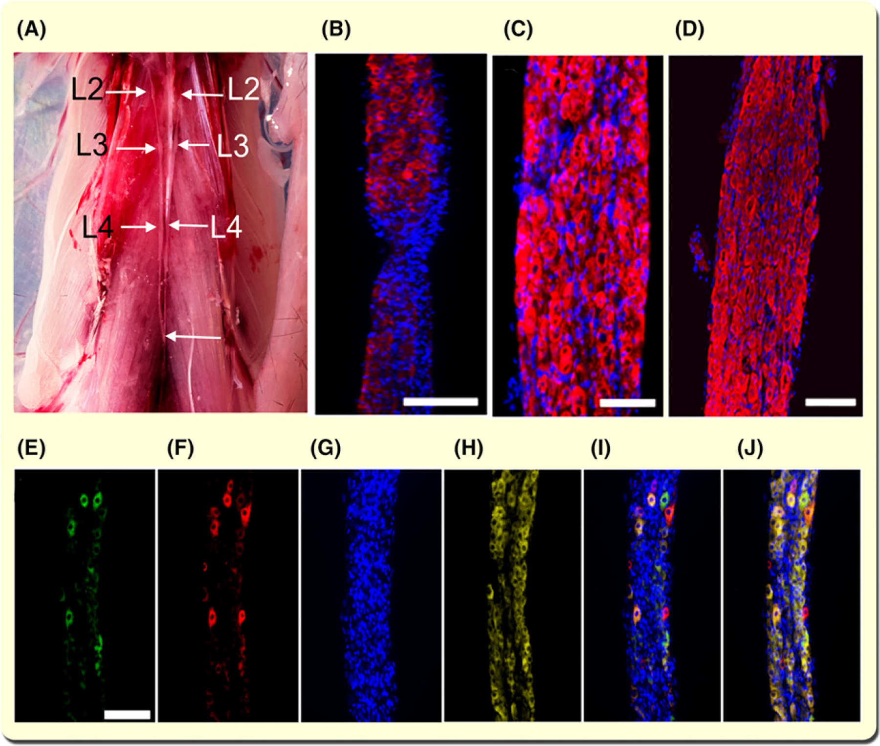
FIGURE 1.

Lumbar sympathetic ganglia establish a functional connection with hindlimb muscles. A, Paravertebral mouse sympathetic ganglia. All abdominal organs and the diaphragm have been excised to expose the paravertebral sympathetic ganglia from lumbar levels L2–L4. The bottom arrow shows the caudal joining of the ganglion chains. B-D, Immunostaining with tyrosine hydroxylase (TH) antibody and staining for Hoechst 33342 (nuclei) of the paravertebral sympathetic ganglia segments. Calibration bar = 500 and 50 μm for B and C respectively. D, Lumbar sympathetic ganglia immunostained with TH antibody, stained with Hoechst 33342, and visualized with confocal microscopy. Z-stacks were generated by scanning 20 contiguous optical slices of 1.16 μm/slice. Calibration bar = 100 μm. E-J, CTB-AF488 and -AF555 fluorescence in lumbar sympathetic ganglia 24 h after their injection into the TA and GA muscles. Images are representative of three experiments. Neuronal cell bodies in the sympathetic ganglia innervating GA (E) and TA (F) are stained with Hoechst 33342 (G) and immunostained with TH (H). Overlay image of E-G is shown in I. Overlay image of E-H is shown in J (bar = 100 μm)
