
FIGURE 4.
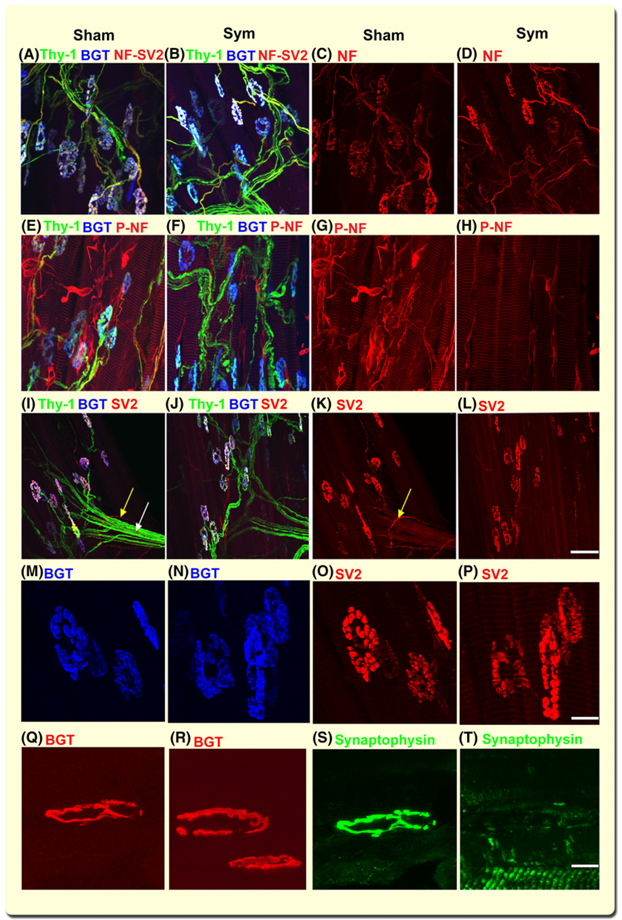
SNS ablation evokes broad neurofilament dephosphorylation and depletion of synaptic vesicles in sympathectomized but not sham mice. Representative z-stack confocal images of lumbricalis muscle neuromuscular junction (NMJ) innervation from sham (A, C, E, and G) and sympathectomized (B, D, F, and H) thy-1 transgenic mice. Thy-1+ motor axonal terminals (green) and BGT-680 (blue, cy5.5) (A, B) co-registered with nonphosphorylated NF antibody (red, AF568-SMI 311 Ab) (C, D) in sham (A, C) and sympathectomized (B, D) mice. Thy-1+ axons overlap with phosphorylated NF (AF568-SMI 312 Ab) axons in sham (E, G) but not sympathectomized (F, H) mice. N = 14 random images from six lumbricalis muscles, five mice per group. Representative 2D z-stack confocal images of motor axons immunostained with SV2 antibody (red, AF568) and postterminal stained with BGT-680 (blue, cy5.5) in lumbricalis muscles from sham (I, K) and surgically sympathectomized (J, L) thy-1 (green) transgenic mice. SV2 immunostaining outlines the vascular (yellow arrow) and axonal (white arrows) trajectory in sham (K) but not sympathectomized (l) mice. Notice that axons and their terminals are thy-1+ in J and SV2 outlines axonal terminals in sham (K) and sympathectomized (l) mice. N = 8 confocal fields analysed in four sham and four sympathectomized mice. Bar = 50 μm. M-P are close-ups of the insets in i-l, displaying the postterminals stained with BGT (blue) and immunostained for SV2 (red). Calibration bar = 10 μm. Q-T show representative z-stack confocal images of NMJ postterminals stained with BGT-568 (red) and axon terminals immunostained with synaptophysin antibody (green, AF488) in lumbricalis muscles from sham and surgically sympathectomized mice. Calibration bar = 10 μm