. 2020 Apr 20;9:e54304. doi: 10.7554/eLife.54304

Figure 2. Teasing out a WNT-sensitive molecular signature based upon transcriptome profiling of skin progenitors possessing different WNT signaling levels.

(A) LV construct, epifluorescence imaging and FACS strategy for isolating WNT signaling (GFP+) and WNTlo skin progenitors from LV-transduced E14.5 Apc fl/+ and Apcfl/fl; R26fl-stop-fl-tdTOM embryos. (B) Gene set enrichment analysis (GSEA) of gene sets showing marked differential expression in WNT signaling progenitors from Apc-null vs Apc-het embryos. False discovery rate (FDR) q-values of enrichment are shown for each gene set. (C) Waterfall plot depicting genes markedly influenced (Log2 Fold Change ≥ 1.5, p<0.05) by APC status (color-coding according to B). (D) Volcano plot showing differentially regulated transcripts and WNT-reporter status.

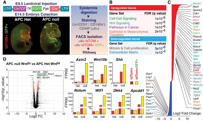
Figure 2—source data 1. Source data for the graphs shown in Figure 2D.

Figure 2.

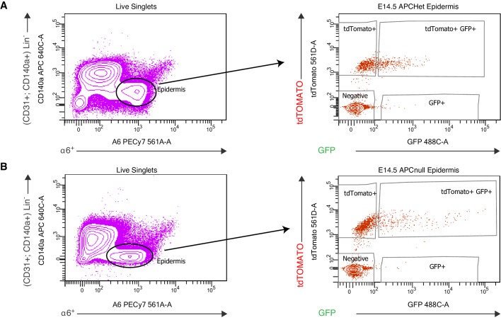
Figure 2—figure supplement 1. FACS purification strategy to isolate WNThi skin progenitors from Apc embryonic skin.

Figure 2—figure supplement 1.

(A–B) FACS purification of WNThi cells from Apcfl/+; Rosa26-lox-stop-lox-dtTomato embryos (A) and from Apcfl/fl Rosa26-lox-stop-lox-dtTomato embryos (B). Both types of embryos were transduced at E9.5 with an LV harboring a WNT GFP reporter and Pgk-Cre and then harvested at E14.5. WNT-reporterhi (a6+ tdTomato+ GFP+) and WNT-reporterlo (a6+tdTomato+) progenitors were purified, while suprabasal (a6neg tdTomato+ GFPneg) epidermal cells and non-epidermal cells (Lin-: CD131+ and CD140a+) were eliminated.
Figure 2—figure supplement 2. Cell adhesion transcripts upregulated in Apc null WNThi cells (Geneontology – PANTHER Classification System).

Figure 2—figure supplement 2.

Figure 2—figure supplement 2—source data 1. Cell adhesion transcripts upregulated in Apc null WNThi cells (Geneontology – PANTHER Classification System).