. 2020 Apr 20;9:e54304. doi: 10.7554/eLife.54304

Figure 5. Evidence of oppositely polarized and short-range action of WNTs and WNT inhibitors in hair bud progenitors that are actively signaling through WNTs.

(A) LV-constructs and strategy to monitor WNT inhibitors and WNTs. M, myc-tag. TRE, tetracycline regulatory element. Krt14rtTA is a transgenic mouse line expressing a doxycycline (DOX)-inducible transcriptional activator for TRE. (B, C) Similar to endogenous expression, anti-WIF1 and anti-NOTUM immunofluorescence on MYC-tag transduced skin show apical localization in hair bud progenitors. (D) Anti-MYC-tag immunofluorescence of transduced skins revealing apical polarization of NOTUM and WIF1, but basal polarization of WNT10B. At right are basal-apical MYC-Tag/DAPI pixel intensity profiles of basal hair bud progenitors (n = ≥40 WNT signaling progenitors; mean ± SEM; a.u., arbitrary units). (E) Pixel intensity profile and immunolocalization of endogenous WNT-receptor FRIZZLED 10 shows uniform localization at borders of hair bud and germ WNT signaling cells (n = 37 cells; mean ± SEM; a.u., arbitrary units). *Denotes magnified cells, shown at right of each frame. Scale bars: 5 μm magnified cells; all the others, 20 μm.

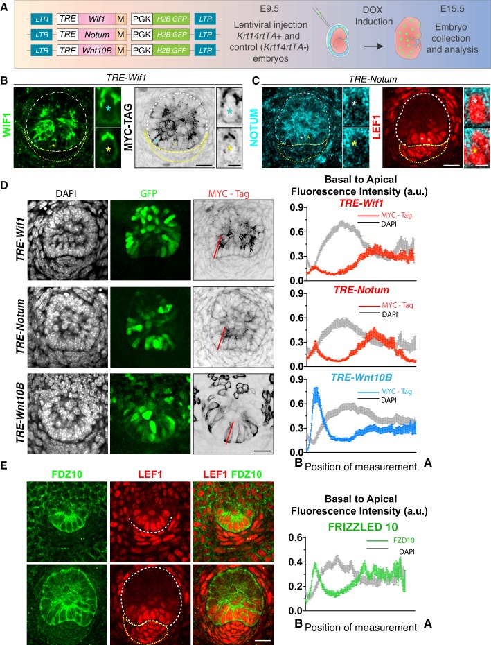
Figure 5—source data 1. Source data for the graphs shown in Figure 5D and E.
elife-54304-fig5-data1.xlsx (944.6KB, xlsx)

Figure 5.

Figure 5—figure supplement 1. WIF1 expression during early hair follicle development.

Figure 5—figure supplement 1.

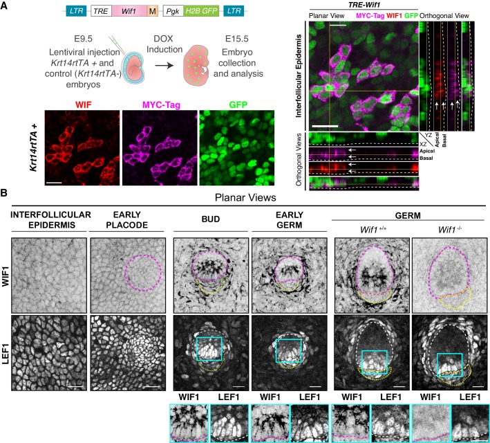
(A) Strategy to monitor WIF1 expression by MYC-tagging (M). Depicted LV was introduced into the amniotic cavities of E9.5 Krt14rtTA embryos, and transduced genes were activated by doxycycline (DOX). Representative whole-mount and orthogonal views of WIF1-MYC-tag and WIF1 immunostaining of E15.5 embryos. Note uniform distribution of protein within the plane of the embryonic epidermis (white arrows). Dashed line demarcates the border between epidermis and dermis. Yellow lines represent the corresponding orthogonal views. Scale bar, 20 μm. (B) Endogenous WIF1 localization is shown by whole-mount immunofluorescence of interfollicular epidermis, hair placodes, hair buds and hair germs. Note that WIF1 is increased in the hair bud and germ epidermis but is only expressed at the bud stage in the early dermal condensate (yellow dotted lines). Note also the distinct apical localization of WIF1 in hair buds and germs. Wif1 full KO embryos were used to test the specificity of the antibody, shown here for the hair germ stage. Boxed areas in blue are magnified below each image, with WIF1 and LEF1 immunolabels. Scale bars, 20 μm.
Figure 5—figure supplement 2. NOTUM expression during early hair follicle development.

Figure 5—figure supplement 2.

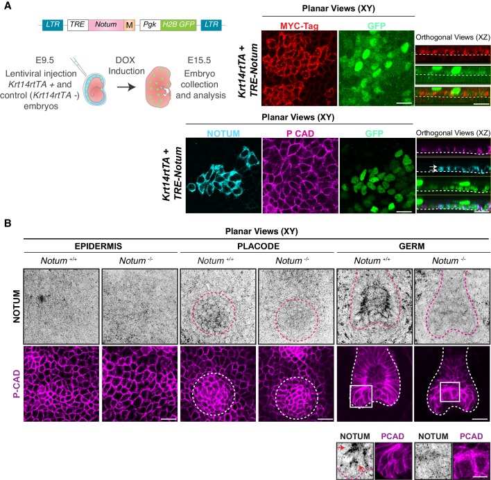
(A) Strategy to monitor NOTUM by MYC-epitope tagging (M). Depicted LV was introduced into the amniotic cavities of E9.5 Krt14rtTA embryos and the transduced Notum gene was activated by doxycycline (DOX). Representative whole-mount planar (XY) and orthogonal (XZ) views of NOTUM-MYC-tag immunostaining. Note that within the plane of embryonic epidermis, NOTUM is uniformly distributed (white arrows). Dashed lines demarcate the border between epidermis and dermis. (B) Endogenous NOTUM immunolocalization in wild-type and Notum-null skin, is shown by whole-mount immunofluorescence of interfollicular epidermis, hair placode and hair germs. P-Cadherin co-immunolabeling was used to mark the skin epithelium. Dotted circles/lines encase the developing hair follicles. Boxed areas are magnified in the boxes below the relevant images. Note little or no NOTUM in interfollicular epidermis. However, NOTUM is expressed in developing hair placodes. At the hair germ stage, it is clear that endogenous NOTUM protein concentrates on the apical side of WNThi epithelial cells (insets, red arrows). Notum-null skin was used as a control to show the specificity of the NOTUM antibody. All scale bars, 20 μm.
Figure 5—figure supplement 3. Differential polarization of WNT inhibitors and activators.

Figure 5—figure supplement 3.

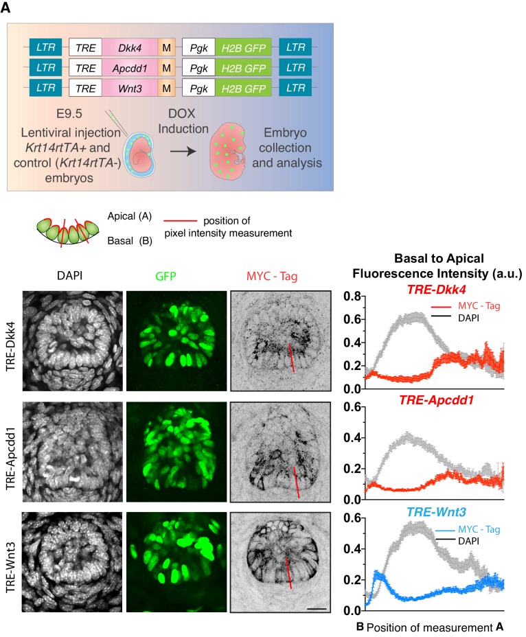
Strategy to monitor WNT inhibitors (DKK4 and APCDD1) and ligands (WNT3) by MYC-epitope tagging (M). LV-constructs were introduced into the amniotic cavities of E9.5 Krt14rtTA embryos, and transduced genes were activated by doxycycline (DOX). Whole-mount immunofluorescence and/or immunohistochemistry of representative images of hair buds are shown below. DAPI to label chromatin; anti-GFP to mark nuclei of transduced cells; Anti-MYC-tag to label the expressed inhibitor/ligand. At right are shown the basal-apical MYC-Tag pixel intensity profiles, which were measured along the lines presented in the overlying schematic of the WNThi progenitor cells of the hair follicle. A minimum of 40 WNT-signaling progenitor cells were analyzed and averaged to develop these profiles. Note the preferential apical enrichment of DKK4 and APCDD1 and opposing basal enrichment of WNT3 signal. Mean ± SEM. a.u., arbitrary units. All scale bars 20 μm.
Figure 5—figure supplement 3—source data 1. Source data for the graphs shown in Figure 5—figure supplement 3A.