
Fig. 1.
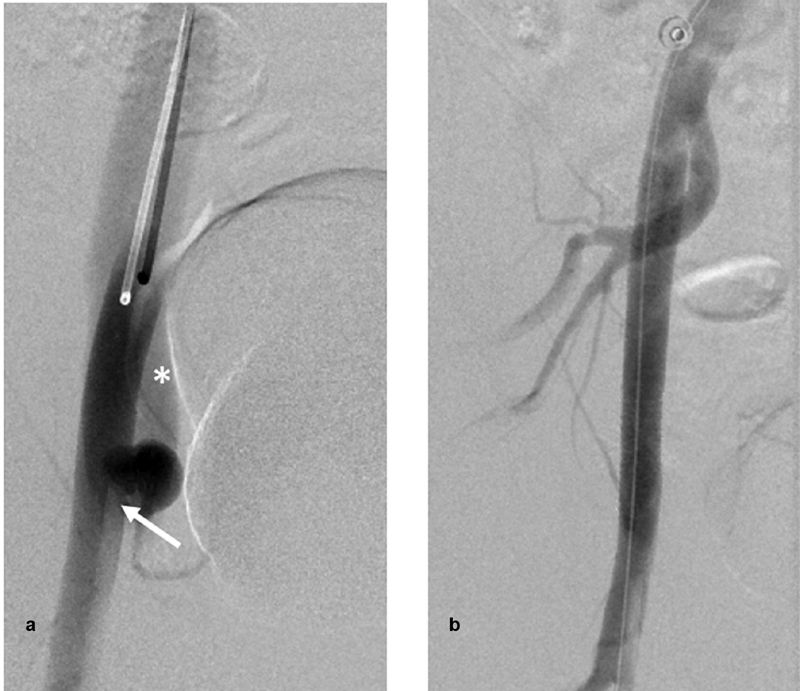
( a ) Digital subtraction angiography (DSA) of the right external iliac artery (EIA) via a left femoral approach anteroposterior view showing a pseudoaneurysm adjacent to the distal tip of the sheath (arrow) with early opacification of the external iliac vein, consistent with arteriovenous fistula (asterisk). ( b ) DSA of the right EIA revealing complete obliteration of aneurysm and the fistula after deploying a Viabahn Gore covered stent across the neck of the aneurysm.