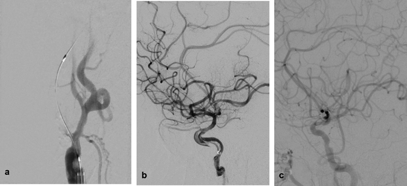
Fig. 2.

( a ) Digital subtraction angiography (DSA) of the right common carotid artery, right anterior oblique view showing total internal carotid artery (ICA) origin occlusion after microcatheter and a wire were successfully advanced through the occlusion. ( b ) DSA run of the right ICA, lateral view after reperfusion of the intracranial vessels showing a linear/spiral filling defect within the petrocavernous segment of the ICA consistent of dissection. ( c ) Repeat angiography 7 days after the procedure showing complete resolution of the dissection with preserved antegrade flow.
