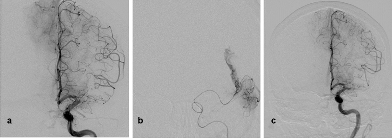
Fig. 3.

( a ) Digital subtraction angiography (DSA) of the left internal carotid artery (ICA), Towne view showing occluded left middle cerebral artery (MCA) with anterior cerebral artery pial collaterals retrogradely filling the distal MCA territory. ( b ) Microcatheter DSA run of the MCA after first pass using stent retrieval device (Solitaire) revealing contrast extravasation at the distal MCA. ( c ) Guide catheter DSA run of the left ICA showing occluded MCA sealing the microperforation. The presence of collaterals suggests acute on top of chronic occlusion or long-term MCA stenosis.
