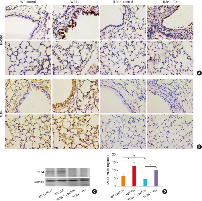
Fig. 4. TLR4 deficiency did not affect HMGB1 production in TDI-treated mice. (A) Representative immunohistochemistry of HMGB1 in the bronchial regions (upper panel) and alveolar regions (lower panel). Original magnification was 400×. (B) Representative immunohistochemical staining of TLR4 in the bronchial regions (upper panel) and alveolar regions (lower panel). (C) Protein expression of TLR4, HMGB1 in lung homogenates were detected by Western blotting (n = 6). (D) Levels of HMGB1 in BALF of different groups were detected by ELISA (n = 4–6).
WT, wild-type; TLR4, toll-like receptor 4; TDI, toluene diisocyanate; NS, not significant; HMGB1, high mobility group box 1; BALF, bronchoalveolar lavage fluid; GAPDH, glyceraldehyde 3-phosphate dehydrogenase; ELISA, enzyme-linked immunosorbent assay.
*P < 0.05.

