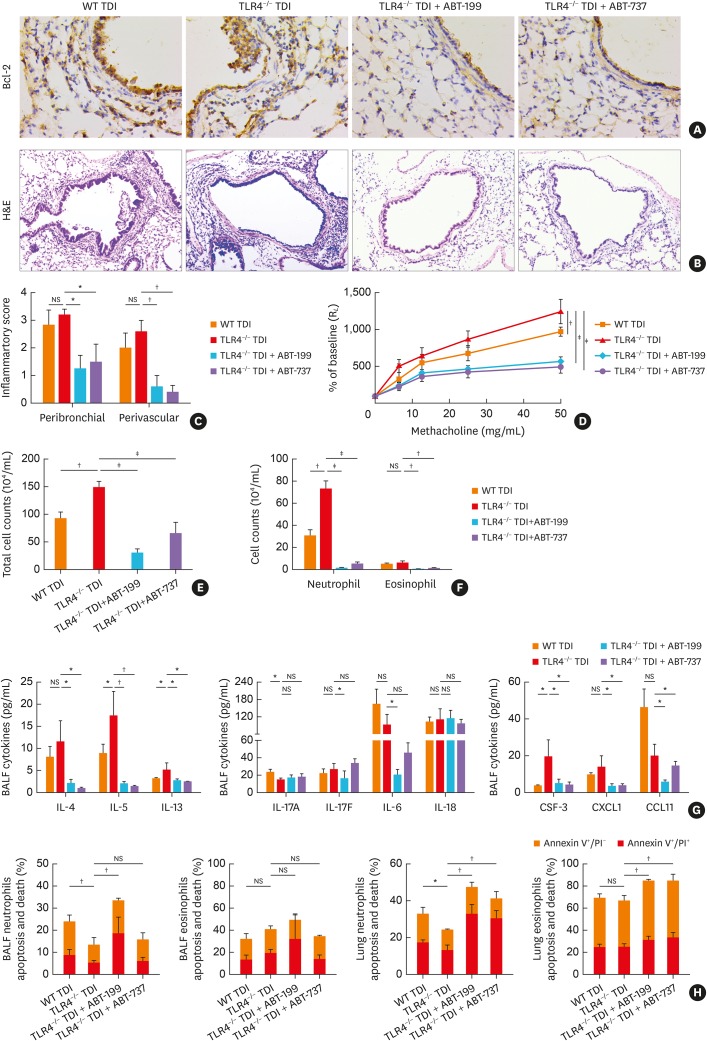
Fig. 6. Bcl-2 inhibitors alleviated the aggravated airway inflammation in TDI-exposed TLR4–/– mice. (A) Representative immunohistochemistry of Bcl-2 in the bronchial regions. Original magnification was 400×. (B) Representative H&E-stained lung sections of different groups. Original magnification was 200×. (C) Semi-quantification of peribronchial and perivascular inflammation was performed (n = 5-6). (D) Airway hyperresponsiveness was measured by RL. Results were shown as percentage of baseline (n = 4). (E) Numbers of total inflammatory cell in BALF (n = 5–6). (F) Numbers of neutrophils, and eosinophils in BALF (n = 5–6). (G) Levels of Th2-related cytokines (IL-4, IL-5 and IL-13), Th17-related cytokines (IL-17A, IL-17F, IL-6 and IL-18), eosinophil chemoattractant CCL11, and neutrophil chemoattractant CSF-3, CXCL1 in BALF (n = 4–6). (H) Numbers of apoptotic ex vivo eosinophils and neutrophils were determined.
WT, wild-type; TLR4, toll-like receptor 4; TDI, toluene diisocyanate; NS, not significant; RL, lung resistance; Bcl-2, B-cell lymphoma 2; IL, interleukin; CCL11, C-C motif chemokine 11; CXCL1, chemokine (C-X-C motif) ligand 1; CSF-3, colony-stimulating factor 3; BALF, bronchoalveolar lavage fluid; Th, T helper; H&E, hematoxylin and eosin; PI, propidium iodide.
*P < 0.05; †P < 0.01; ‡P < 0.001.

