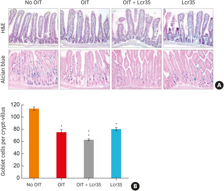
Fig. 4. Histological findings of the small intestine of mice treated with egg OIT and Lcr35. (A) Hematoxylin and eosin stain (upper, ×200) and Alcian blue stain (lower, ×400). (B) Mucus cell count in the small intestine.
OIT, oral immunotherapy.
*P < 0.05, compared to the control group; †P < 0.05 compared to the OIT group.

