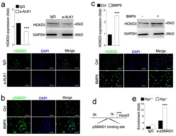
Figure 3. BMP9 induced the expression of HOXD3 through the ALK1 and pSMAD1.
(a) Expression of HOXD3 in HUVECs treated by anti-ALK1 neutralizing antibodies as shown by real-time PCR, immunoblotting and immunofluorescence staining.
(b) Level of pSMAD1 in HUVECs treated with BMP9 as shown by immunostaining.
(c) Induction of HOXD3 in HUVECs treated by BMP9 as shown by real-time PCR, immunoblotting and immunostaining.
(d) pSMAD1 DNA-binding site in the promoter of the Hoxd3 gene.
(e) Increased pSMAD1 DNA-binding at the Hoxd3 promoter in Mgp−/− aortas as shown by ChIP-assays.
Data were analyzed by Student’s t test. ***, P<0.001. Error bars are standard deviation. ***, P < 0.001. Scale bar, 50 μm.

