Figure 5:
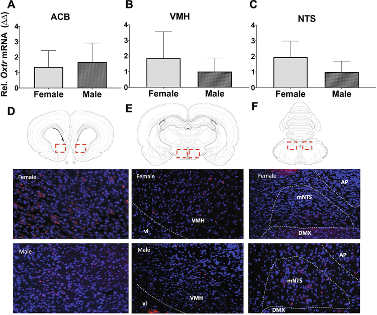
Expression of Oxtr in feeding-relevant neural sites. Relative Oxtr mRNA expression (top panels) in tissue punches from the nucleus accumbens (ACB; a), ventromedial hypothalamus (VMH; b), and nucleus tractus solitarius (NTS; c) from male and female rat brains. Representative images of coronal brain sections (bottom panels) showing fluorescence in situ hybridization for Oxtr (red) in the ACB (a), VMH (b), and NTS (c) with dapi counterstain (blue). Data are means ± SEM.

