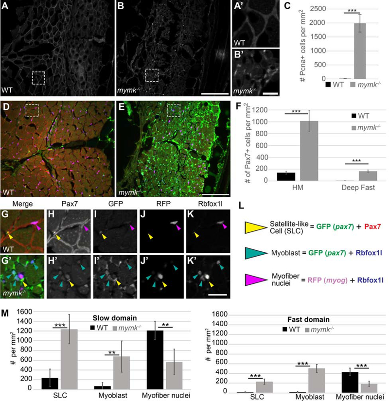
Figure 6: Cell proliferation and Pax7-positive satellite-like cell number are dramatically increased in adult mymk mutants.
(A, B) Transverse sections near the horizontal myoseptum (HM) of wild-type (WT) (A) and mymk mutant (B) adult skeletal muscle showing Pcna expression (grey) which marks proliferating nuclei. (A’ and B’) Magnified images of boxed regions in A and B. (C) Graph showing the average number of Pcna-positive cells per mm2 near the HM in WT and mymk mutant adults. (D, E) Transverse sections of pax7:GFP (green); myog:H2B-mRFP (magenta) transgenic WT (D) and mymk mutant (E) adult skeletal muscle colabeled for Pax7 (red), Rbfoxll (blue), and pax7a:GFP (green). pax7a:GFP marks satellite-like cells (SLCs) as well as cells that have begun to differentiate; SLCs can be unambiguously identified by their intensely Pax7-positive nuclei. myog:H2B-mRFP (magenta) marks myonuclei; expression of myog:H2B-mRFP and pax7:GFP transgenes rarely overlap in adult WT muscle (Berberoglu et al., 2017). (F) The average number of Pax7-positive cells per mm2 near the HM and in dorsal fast muscle of WT and mymk mutant adults. (G-K’) Magnified views of boxed regions in D and E show the merged (G, G’) and individual confocal channels for Pax7 (H, H’), pax7a:GFP (I, I’), myog:H2B-mRFP (J, J’) and Rbfoxll (K, K’) in adult WT (G-K) and mymk mutant (G’-K’) muscle. Arrowheads indicate SLCs (yellow), myoblasts (teal), and myofibers (magenta). (L) Summary of cell types based on expression overlap. (M) Number of SLCs (strongly Pax7-positive and GFP-positive), myoblasts (GFP-and Rbfoxll-positive), and myofibers (Rbfoxll-and RFP-positive) in WT and mymk mutant adult slow and fast muscle domains. Student’s f-test (p*** < 0.001, p** < 0.01). Scale bar in B (for A and B) and E (for D, E) is 100 μm, in B’ (for A’ and B’) is 15 μm, and in K’ (for G-K’) is 20 μm.

