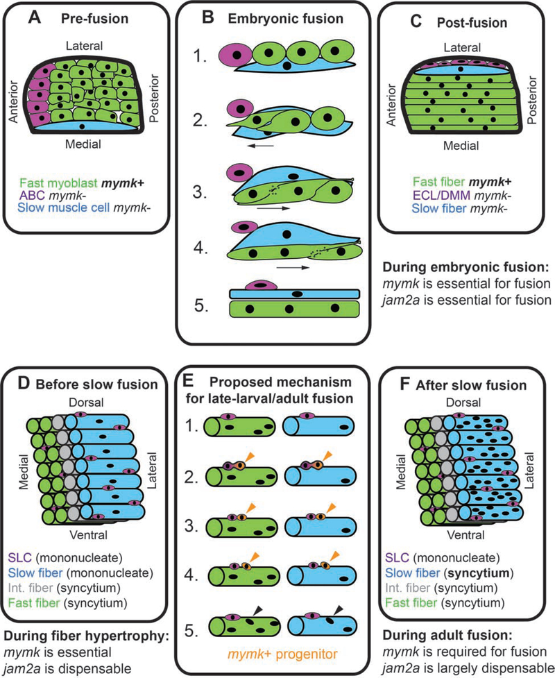
Figure 9: Muscle fusion mechanisms are developmentally regulated.
Model describing developmentally-regulated muscle fusion in zebrafish. (A) Diagram of cell positions in a newly formed somite, viewed from dorsal, showing anterior border cells (ABCs; purple), fast myoblasts (green), and an elongated slow muscle cell (blue). (B) Cell dynamics during embryonic cell fusion. (B1, B2) As a slow muscle cell moves laterally, ABCs also shift position and are replaced at the anterior border by fast myoblasts. (B3-B5) Fast myoblasts then sequentially fuse toward the posterior somite border. (C) Diagram of a somite after slow muscle cell migration. At this stage, slow muscle cells are mononucleate and fast muscle cells are multinucleate. After shifting position, ABCs are called the external cell layer (ECL) in some studies and dermomyotome (DMM) in others. See reviews for further details on embryonic muscle development in zebrafish (Goody et al., 2017; Jackson and Ingham, 2013; Li et al., 2017; Sampath et al., 2018; Stellabotte and Devoto, 2007). (D) Muscle arrangement in late larval stages, prior to slow muscle fusion. By this stage there are three fiber types: fast (green), intermediate (int., gray), and slow (blue), and the slow muscle region is enriched for satellite-like cells (SLCs, purple) (Berberoglu et al., 2017). (E) In late-larval stages, we hypothesize that SLCs produce mymk-positive muscle progenitors (orange). These progenitors could potentially fuse into both fast and slow muscle fibers (orange arrowheads), resulting in the addition of new myonuclei (black arrowheads). (F) By late-larval stages, all muscle fiber types are multinucleate.

