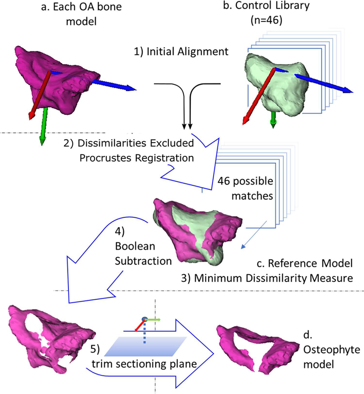
Fig. 2.
Flow chart of the five main steps in generating osteophyte bone models for the trapezium. Steps for the first metacarpal were identical. 1) The articular coordinate systems of the bone models were computed (a and b) and used as an initial registration seed for all 46 control models (b) to each bone model with osteoarthritis (OA). 2) Procrustes superimposition was used to attain the final registrations. 3) The control bone with the minimum dissimilarity measure was identified as that OA model’s reference bone (c). 4) Osteophyte models were calculated by Boolean subtraction of the reference bone model from the OA model. 5) The osteophytes were then trimmed by a sectioning plane to generate the osteophyte bone model relevant to the first carpometacarpal joint (d).

