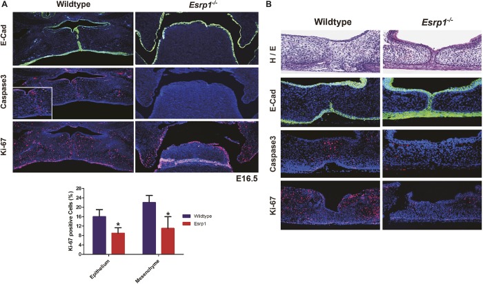
Fig. 3.
Reduced proliferation and a fusion defect contribute to cleft palate in Esrp1−/− mice. (A) Coronal sections stained for E-cadherin (green) in E16.5 control and Esrp1−/− embryos showing the medial edge epithelial seam (MES) becoming discontinuous following fusion in controls whereas palatal shelves failed to elevate in the mutants. Activated caspase 3 staining shows apoptosis at the site of fusion, but no apparent increase in apoptosis in Esrp1−/− embryos, which also show reduced Ki-67 staining for proliferating cells compared with WT in the palatal shelves. Inset shows higher magnification of caspase 3 staining at the site of fusion. Graph shows quantification of the percentage of Ki-67-positive cells out of total cells in WT (n=3) and Esrp1−/− embryos (n=3). Error bars indicate s.d. Statistical significance was determined by two-tailed t-test. *P<0.05. Error bars indicate s.d. (B) Palatal organ culture showing lack of dissolution of the MES and associated apoptosis and reduced proliferation in palatal shelves from Esrp1−/− embryos compared with WT.

