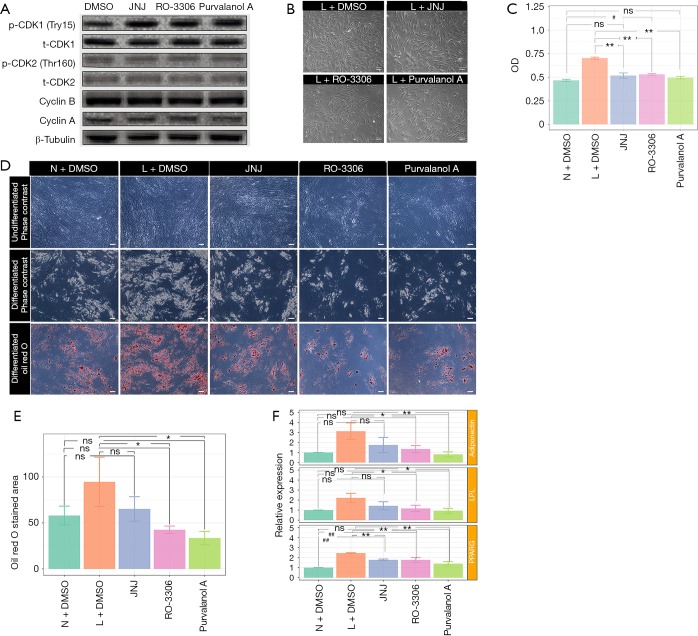
Figure 6.
Cell cycle inhibitor influences the proliferative ability and differentiation potential of ASCs. (A) Immunoblot analysis of p-CDK1, CDK1, p-CDK2, CDK2, Cyclin B and Cyclin A expression levels after the cells were treated or not treated with CDK inhibitors JNJ, Ro-3306, or purvalanol A for 2 h; (B) representative photos of cell density and morphology of ASCs after treatment with CDK inhibitors JNJ, Ro-3306, or purvalanol A for 48 h. Bar =100 µm; (C) the division potential of ASCs was detected with CCK8 after the cells were treated or not treated with CDK inhibitors JNJ, Ro-3306, or purvalanol A for 48 h. Values were expressed as the mean ± SEM of three replicates. **, P<0.01, significantly different from L+DMSO; #, P<0.01, significantly different from N+DMSO; ns, no statistical difference (one-way ANOVA). (D) ASCs treated with or without CDK inhibitors, JNJ, Ro-3306, or purvalanol A were induced to differentiate into adipocytes and were stained with Oil Red O; 10 images of each condition were captured for statistical analysis; (E) the areas stained with Oil Red O was expressed as the mean ± SEM (n=3). Bar=100 µm. *, P<0.05, **, P<0.01, significantly different from L+DMSO; ##, P<0.01, significantly different from N+DMSO; ns, no statistical difference (one-way ANOVA); (F) RT-qPCR detected the expression of mature adipocyte markers adiponectin, LPL, or PPARG in induced ASCs, which were treated with CDK inhibitors before adipogenesis. ASCs derived from lymphedema group and normal group of 3 random donors in triplicates. *, P<0.05, **, P<0.01, significantly different from L+DMSO; #, P<0.05, ##, P<0.01, significantly different from N+DMSO; ns, no statistical difference (one-way ANOVA). N, normal; L, lymphedema; ASCs, adipose-derived mesenchymal stem cells; RT-qPCR, quantitative reverse transcriptase-PCR; CDK, cyclin-dependent kinase; CCK8, cell counting kit-8.

