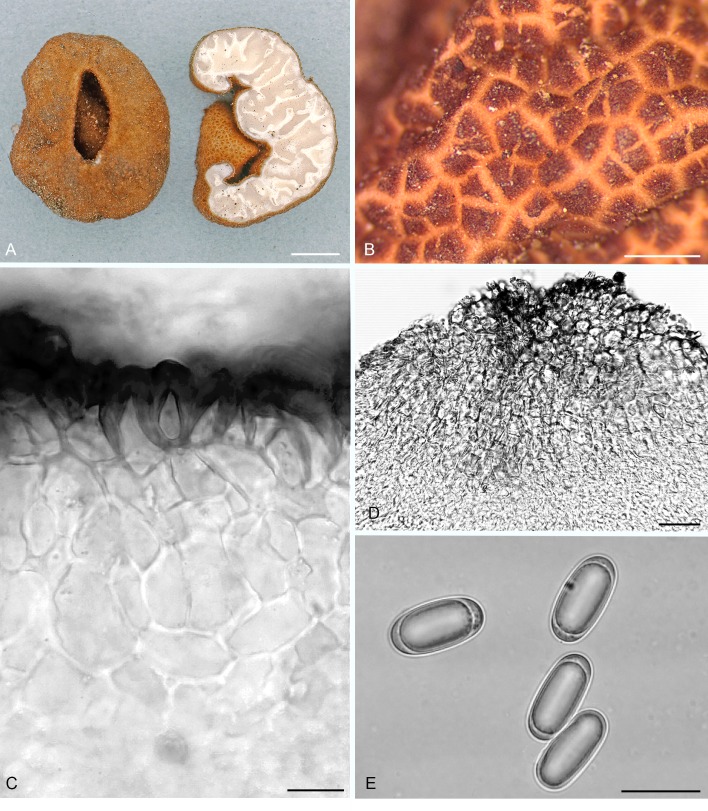
Fig. 10.
Balsamia setchellii. A. Ascoma showing peridial surface, gleba, and peridium in section (OSC 79995). B. Peridial surface showing wart pattern in surface view (OSC 131295). C. Wart structure showing thick-walled cells. D. Cross-section of peridial surface showing wart structure (isotype; OSC 130978). E. Ascospores (OSC 131295). Bars A = 5 mm, B = 750 μm, C = 20 μm, D = 30 μm, F = 20 μm.

