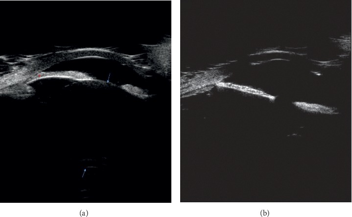
Figure 3.

Ultrasound biomicroscopy examinations of the right and left eyes. Both eyes had a history of retinopathy of prematurity treated with panretinal photocoagulation. (a) The right eye was phakic, and the angle is closed. (b) The left eye was aphakic, and the angle is open. Arrows show anterior and posterior lens capsules; asterisks show iridocorneal adhesion.
