Figure 1.
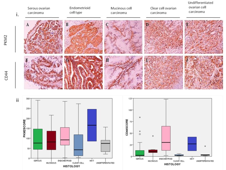
(i) Immunohistochemical (IHC) representative staining of PKM2 in the cytoplasm and of CD44 mainly in cell membranous and in cytoplasm in different histological subtypes of EOC tumor tissues (magnification 200×). (A) PKM2 and (F) CD44 staining in serous ovarian carcinoma. (B) PKM2 and (G) CD44 staining in endometroid cell type. (C) PKM2 and (H) CD44 protein expression in mucinous cell carcinoma. (D) PKM2 and (I) CD44 in clear cell ovarian carcinoma. (E) PKM2 and (J) CD44 expression in undifferentiated ovarian cell carcinoma. (ii) Boxplots showing PKM2 and CD44 protein expression distribution by different histology subtypes of ovarian carcinoma specimens (Wilcoxon rank test p-value = 0.004 for CD44 and p-value = 0.540 for PKM2 protein expression). PKM2: pyruvate kinase M2; EOC: epithelial ovarian cancer; *: extreme outliers.

