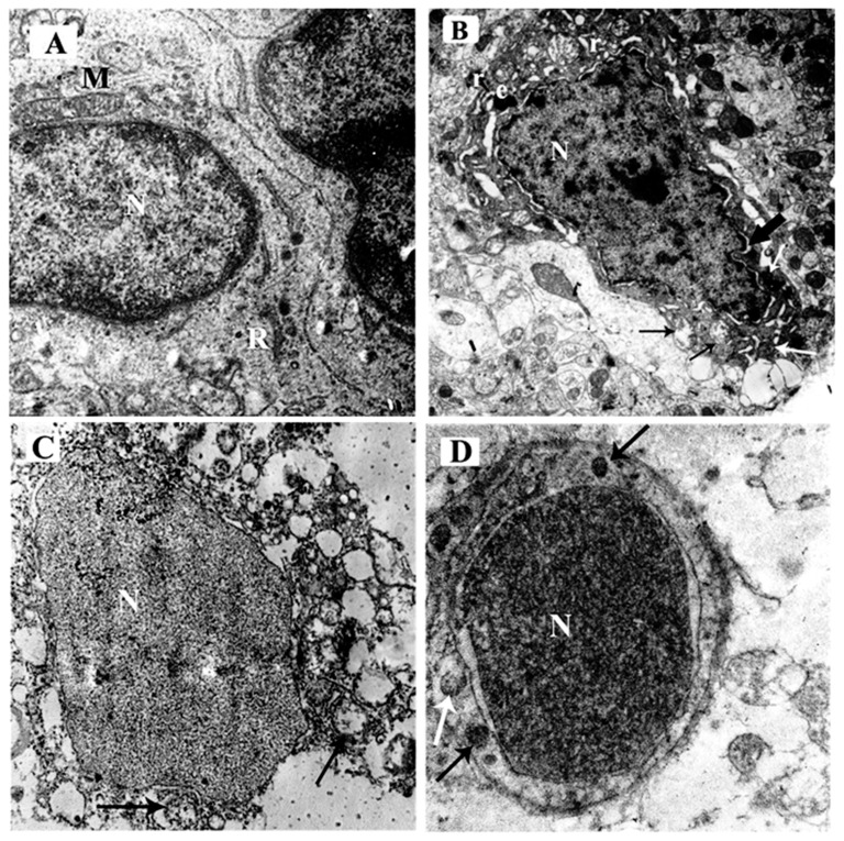
Figure 7.
Electron micrographs of the mouse hippocampus in the CA3 region of the different study groups; (A) control group and ceftriaxone group; the pyramidal cell has a euochromatic, central, and rounded nucleus (N) with a smooth bi-laminar nuclear envelope and fine dispersed chromatin. Well-formed rough endoplasmic reticulum (R) and intact mitochondrial (M) are seen. (B) ARS group demonstrating distorted dense neuron with an irregular outline. The nucleus (N) has marginated chromatin, chromatin clumps, and is surrounded by perinuclear space (thick black arrow). The cytoplasm shows shrinkage and deposition of electron-dense bodies (e). Some mitochondria are distended with disrupted cristae (black arrow) while others are comparable to the control (white arrow). The rough endoplasmic reticulum is markedly dilated (r). (C) ARS group demonstrates markedly affected pyramidal cells with an irregular nucleus (N) and multiple ballooned mitochondria (arrows) with disrupted cristae. (D) ARS + ceftriaxone group; the pyramidal cell has a regular-shaped nucleus (N) and is surrounded by a bi-laminar nuclear membrane. The cytoplasm reveals multiple mitochondria comparable to the control (black arrow) while one is ruptured (white arrow). (× 8000).

