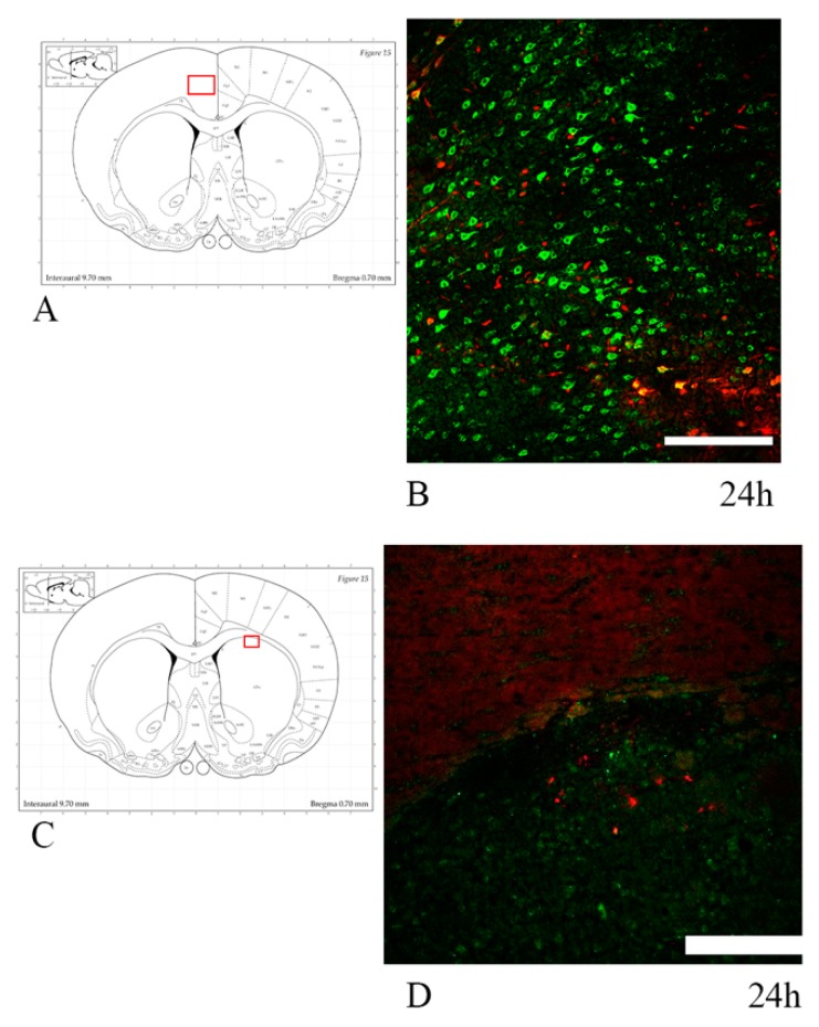
Figure 13.
HSP70/APP double-staining in 24 h trauma-exposed animals. (A,C) The highlighted area refers to the area represented in the adjacent microphotograph. (B) HSP70 and APP expression in the cingulate cortex. Few neurons are positive for both markers. (D) HSP70 and APP expression in the caudoputamen, close to its interface with the corpus callosum. Scale bar = 200 µm.

