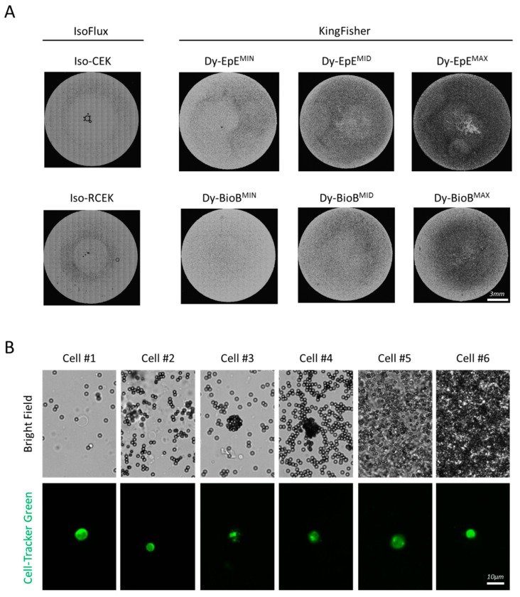
Figure 2.
Identification of enriched pre-labelled cells among the beads. (A) Distribution of Iso-CEK, Iso-RCEK, Dy-EpEMIN,MID,MAX and Dy-BioBMIN,MID,MAX beads in field of a three-field microscope slide used for enumeration of enriched cells. Each image is a montage of all 357 tiled bright field images covering the complete field. (B) Six individual cells identified in one same sample enriched with Dy-EpE beads.

