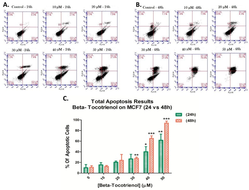
Figure 6.
Beta-Tocotrienol induced apoptosis in MCF7 cells. A dual staining with Annexin V-FITC (FL1-H) and propidium iodide (FL2-H) was assessed to measure the amount of apoptosis of MCF7 cells by flow cytometry, upon 24 h (A) and 48 h (B) of treatment. Results were obtained by C-Flow software. The percentages of total apoptosis were quantified upon beta-T3 treatment and presented in histograms (C). *, ** and *** indicate p < 0.05, p < 0.001 and p < 0.0001 respectively.

