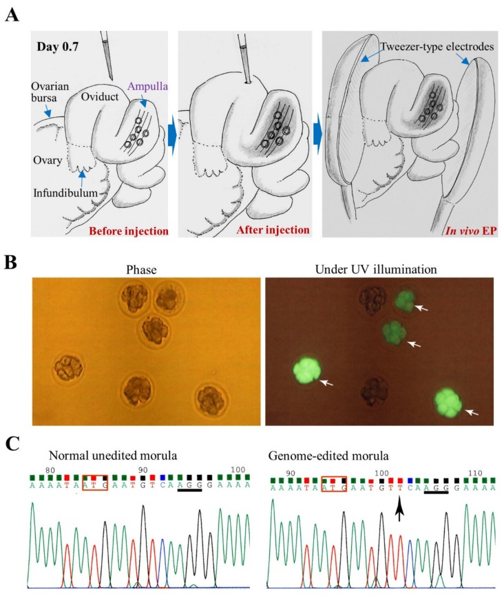
Figure 5.
i-GONAD in mice for targeting GGTA1 locus coding for α-1,3-galactosyltransferase, an enzyme capable of synthesizing cell-surface α-Gal epitope: (A) Schematic illustration of the i-GONAD procedure. Intraoviductal instillation of a solution containing RNP (Cas9/gRNA complex) and fluorescein isothiocyanate (FITC)-labelled dextran 3 kDa (a fluorescent marker for monitoring successful i-GONAD) through the oviductal wall is first carried out on day 0.7 of pregnancy (day 0 of pregnancy is defined as the day a vaginal plug is found). Then, the entire oviduct is subjected to in vivo electroporation (EP) using tweezer-type electrodes. (B) Isolated morulae 2 days after i-GONAD: There are some embryos showing FITC fluorescence (indicated by arrows). (C) Direct sequencing of PCR products derived from an i-GONAD-treated embryo shown in B. The sequences corresponding to the region around ATG (boxed) in GGTA1 in normal unedited and successfully edited embryos are shown. Arrow indicate insertion of nucleotide in front of the protospacer adjacent motif (PAM; underlined).

