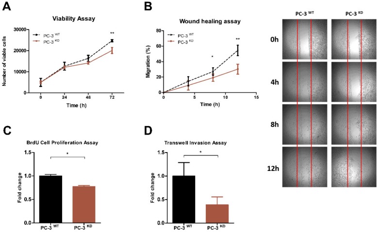
Figure 4.
VIRMA expression promotes PC-3 cells growth and progression in vitro. (A) MTT assay for the number of viable PC-3WT and PC-3KD cells at 24 h, 48 h, and 72 h after seeding. (B) Quantitation of Wound-healing assay for PC-3WT and PC-3KD. Points and connecting line on the left panel represent the migration index of wound-healing assay over the course of 12 h. The distance migrated by PC-3KD cells is represented as relative to that migrated by PC-3WT cells in the same time period. Representative photos are shown in the panel on the right. (C) Proliferation of PC-3WT and PC-3KD cells assessed by BrdU assay at 24 h. (D) Invasion of PC-3WT and PC-3KD cells assessed by Matrigel transwell assay at 24 h. Column bars in (C) and (D) represent the average number of cells from 3 independent experiments. Error bars represent ± SD. ** p < 0.001 * p < 0.05, Mann–Whitney U test.

