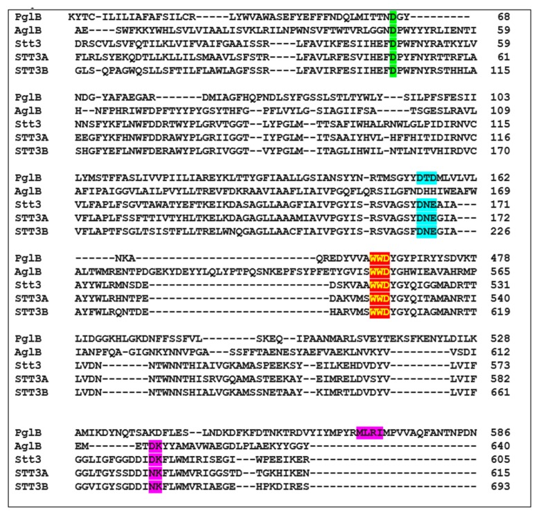
Figure 5.
Sequence alignment of bacterial PglB, archaeon AglB, yeast Stt3, human STT3A, and human STT3B proteins to show the important residues and motifs. D56 (PglB), D47 (AglB and yeast Stt3), D49 (human STT3A), and D103 (human STT3B) are shown in green background. DXD motif in PglB, DXE motifs in yeast Stt3, human STT3A, and human STT3B are shown in cyan background. The conserved WWD motif is shown in red background highlighted in yellow. The MXXI motif in PglB that corresponds to DK motifs in AglB and yeast Stt3 are shown in purple background.

