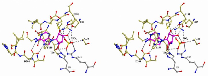
Figure 2.
Structure of the active site of hexameric B. cereus PNP in complex with adenosine and sulfate (PDB 3UAW). The nitrogen, oxygen, and sulfur atoms are shown in blue, red, and yellow, respectively. The carbon atoms of the protein and the substrate are gold and violet, respectively. The atoms of the adjacent subunit of the PNP dimer are colored in grey. The hydrogen bonds involving the adenosine molecule and sulfate are indicated by dashed lines.

