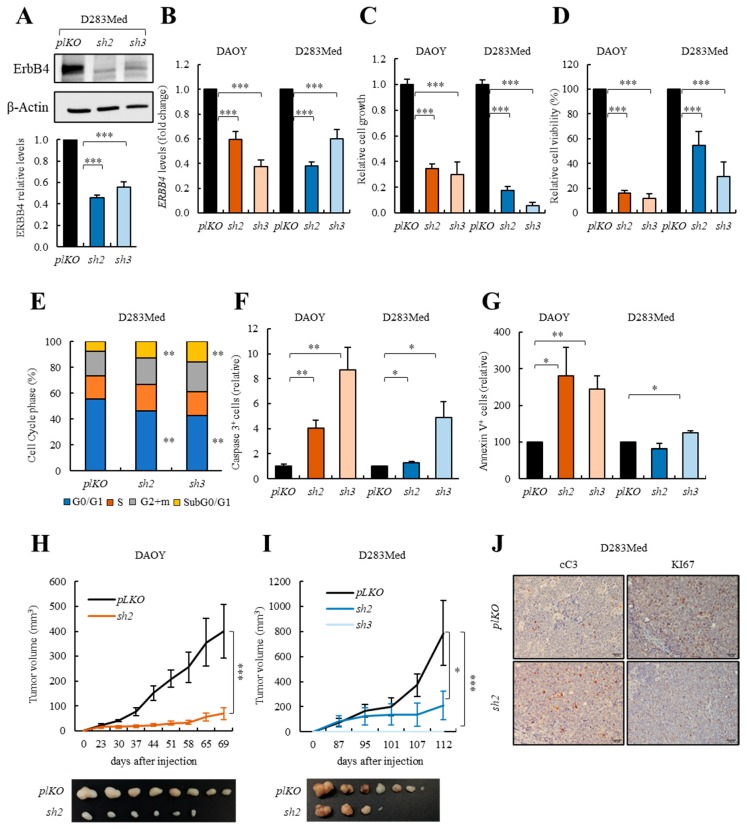
Figure 4.
ERBB4 knock-down impairs medulloblastoma cell viability in vitro and tumor progression in vivo. (A) Representative image and quantification of Western blot analysis of ERBB4 in control (pLKO) and shERBB4 (sh2 and sh3) D283Med cells (n = 4). (B) ERBB4 mRNA expression in control (pLKO) and shERBB4 (sh2 and sh3) cells (n ≥ 9). (C) Relative cell growth at day 5 comparing pLKO with sh2 and sh3 cells (n ≥ 6). (D) MTT studies measuring cell viability in sh2 and sh3 relative to pLKO cells (n ≥ 6). (E) Cell cycle assay measuring the number of cells in each cell cycle phase in pLKO, sh2, and sh3 D283Med cells (n ≥ 3). (F) Immunofluorescence quantification of cleaved-Caspase-3 (cC3) positive cells in sh2 and sh3 relative to pLKO DAOY and D283Med cells (n ≥ 4). (G) Percentage of Annexin-V positive cells in pLKO, sh2 and sh3 DAOY and D283Med cells (n ≥ 3). (H,I) Volume of tumors generated after subcutaneous injection of DAOY and D283Med pLKO, sh2 and sh3 cells (n ≥ 8) at the indicated time points. (J) Representative images of the immunohistochemical staining of cC3 and KI67 in tumors generated after subcutaneous injection of D283Med pLKO and sh2 cells (n = 5). Scale bars = 100 µm. The uncropped blots and molecular weight markers are shown in Figure S11. * p ≤ 0.05; ** p ≤ 0.01; and *** p ≤ 0.001.

