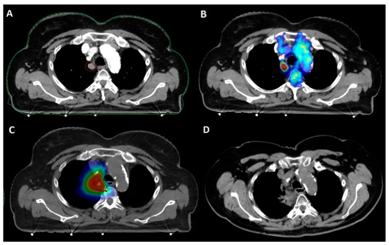
Figure 2.
Patient with a clinical history of pT2N0 lung adenocarcinoma treated with upper lobe lobectomy. A: Paramediastinal middle lobe metastasis (12 × 13 mm) detected at follow-up contrast-enhanced chest CT. B: 18FDG-PET/CT fusion showing isolated hypermetabolism of the paramediastinal metastasis. C: SBRT delivering 50 Gy in five fractions to the PTV (dark blue: 20 Gy; light blue: 30 Gy; green: 40 Gy; yellow: 45 Gy; red: 50 Gy). D: CT evaluation at nine months showing radiation fibrosis following complete metabolic response.

