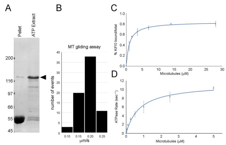
Figure 2.
Motor mechanics. Panel (A) shows microtubule (MT) pellet and ATP extract lanes following the binding and release of the DdKif10725 polypeptide (Coomassie-stained gel, arrowhead denotes position of the Kif10 fusion protein). Panel (B) shows a histogram of MT gliding activity induced by the DdKif10725 fragment, with an average rate of 0.17 μm/s. Panel (C) shows the MT affinity of the motor fragment, plotting % pelleted vs. MT concentration. Panel (D) illustrates motor catalytic activity, plotting the ATPase rate vs. MT concentration. The data in both panels (C) and (D) represent averages from three independent measurements, and error bars indicate standard deviations.

