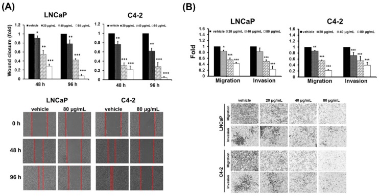
Figure 2.
DFE reduces migration and invasion in PCa cells. (A) A wound healing assay. LNCaP and C4-2 cells were exposed to DFE (20, 40, and 80 µg/mL) or control vehicle. Wound closure was analyzed by the migratory distance at 48 and 96 h, respectively. Results represented as the mean ± SD of three independent experiments. * p < 0.05, ** p < 0.01, *** p < 0.001. Representative images of wound closure in PCa cells exposed to DFE (80 µg/mL) or vehicle at different time points were shown (bottom panel). (B) The migration and invasion of LNCaP and C4-2 cells were determined by the transwell method. The relative migration or invasion was defined as 1.0 (Fold) in the vehicle-treated cells. Data represented the mean ± SD of three independent experiments. * p < 0.05, ** p < 0.01, *** p < 0.001. The images of the migration and invasion of LNCaP and C4-2 cells exposed to vehicle or DFE at 48 h were shown (bottom panel).

