Figure 1.
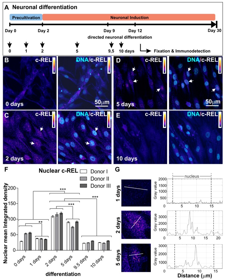
Immunocytochemical analysis of c-REL. (A) Neuronal differentiation procedure. Adult human NCSCs were differentiated into NSCs and further into glutamatergic neurons. NF-κB subunit composition was analyzed at different time points as indicated. (B–E) NCSC-derived NSCs labeled against c-REL after 0, 2, 5 and 10 days of glutamatergic differentiation respectively. Each panel shows c-REL (left) and DNA colocalization (right). Intensity scale indicating white and black as highest and lowest intensity levels. Arrows depict c-REL-nuclear activation. (F) Quantification of immunocytochemical analyses showing c-REL nuclear mean integrated density during early differentiation (n = 3, mean ± SEM). Normality was refuted using Shapiro-Wilk normality test. Nonparametric Kruskal-Wallis (*** p ≤ 0.001) and Bonferroni corrected post-test (*** p < 0.001) revealed significantly increased nuclear translocation of NF-κB-c-REL on days 2 and 5. (G) Fluorescence intensity profiles measured at three different time points (1, 2 and 5 days of differentiation) for cells following transects as shown clearly revealed the difference between nuclear and cytoplasmic fluorescence. NCSCs: neural crest-derived stem cells, NSCs: neural stem cells.

