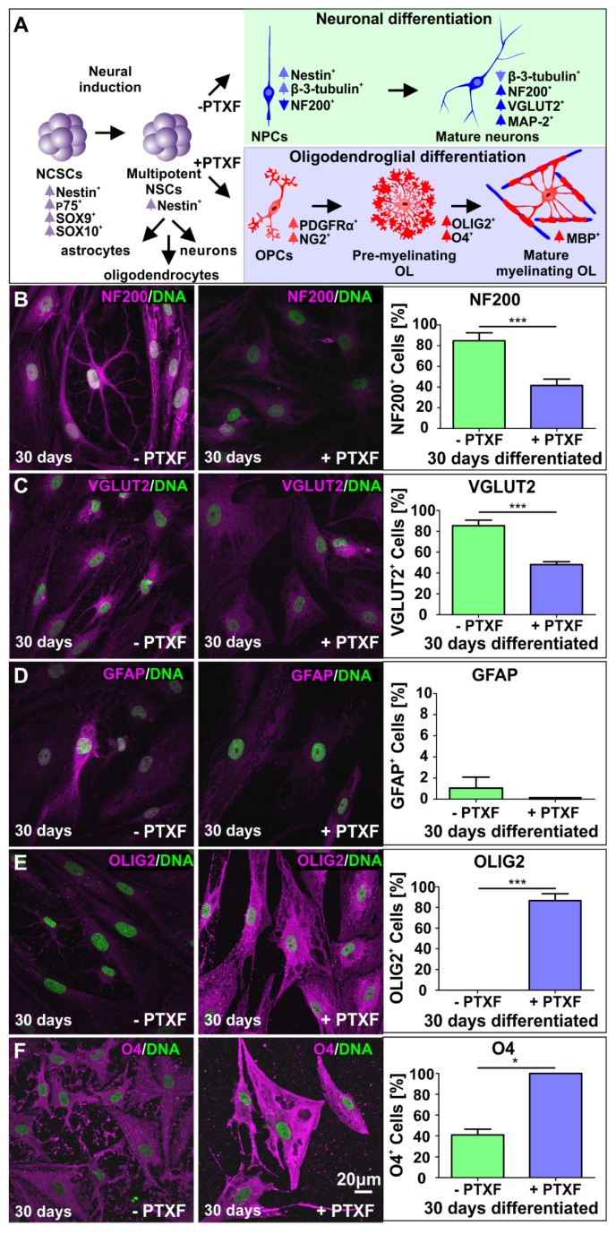
Figure 4.
Immunocytochemical analysis of neuronal and glial markers after glutamatergic differentiation for 30 days in the presence or absence of pentoxifylline. (A) Transcription expression changes during specification of NCSCs into multipotent NSCs with the ability to differentiate into astrocytes, oligodendrocytes and neurons. Differentiation in the absence or presence of PTXF directed the cell fate into the neuronal (light green) or oligodendroglial fate (light blue), further analyzed by the expression of particular markers as indicated (modified from [5,13]). (B) Differentiated NCSC-derived NSCs labeled against NF200 and quantification showing the percentage of NF200+ neurons. Nonparametric Kruskal-Wallis test (*** p < 0.0005) revealed a significant decrease of NF200+ neurons in PTXF-treated cells (41.44% ± 6.31%) compared to untreated neurons (84.68% ± 7.70%). (C) Differentiated NCSC-derived NSCs labeled against VGLUT2, and quantification of VGLUT2+ neurons. Nonparametric Kruskal-Wallis test (*** p < 0.0005) confirmed a significant decrease of VGLUT2+ neurons in the differentiated PTXF-treated cells (47.97% ± 2.72%) compared to untreated neurons (85.37% ± 5.34%). (D) Differentiated NCSC-derived NSCs labeled against GFAP (astrocyte marker) and respective quantified percentage of GFAP+ cells, indicating a very low number of astrocytes present in the untreated control (1.04% ± 1.04%) which are completely absent in the differentiated PTXF-treated cells (0%, nonparametric Kruskal-Wallis test p = 0.3173, no significance). (E) Differentiated NCSC-derived NSCs labeled against OLIG2 (early oligodendrocyte marker) and quantified percentage of OLIG2+ cells, showing no positive cells in untreated control neurons (0%), while most differentiated PTXF-treated-NSCs OLIG2+ (86.67% ± 6.67%). Nonparametric Kruskal-Wallis test (*** p < 0.001) confirmed significant increase of OLIG2+ cells in PTXF-treated differentiated cells, indicating a shift into the oligodendrocyte fate. (F) Differentiated NCSC-derived NSCs labeled against O4 (immature oligodendrocyte marker) and quantification of O4+ cells, showing a significant increase of O4+ cells in PTXF-treated differentiated NSCs (100%) as shown by nonparametric Kruskal-Wallis test (* p < 0.05), compared to untreated neurons (40.97% ± 5.42%). PTXF: pentoxifylline, NCSCs: neural crest-derived stem cells, NSCs: neural stem cells, OPCs: Oligodendrocyte precursor cells, OL: oligodendrocytes, NF200: Neurofilament 200.

