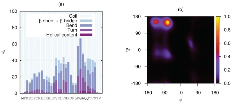
Figure 8.
(a) Stacked secondary-structure histograms per amino acid residue of KEIF as obtained from MD simulations. Three different types of helices included in helical content: (i) -helix, (ii) -helix, and (iii) -helix. This analysis did not include the PPII helix. (b) Ramachandran plot of KEIF as obtained from MD simulations.

