Figure 1.
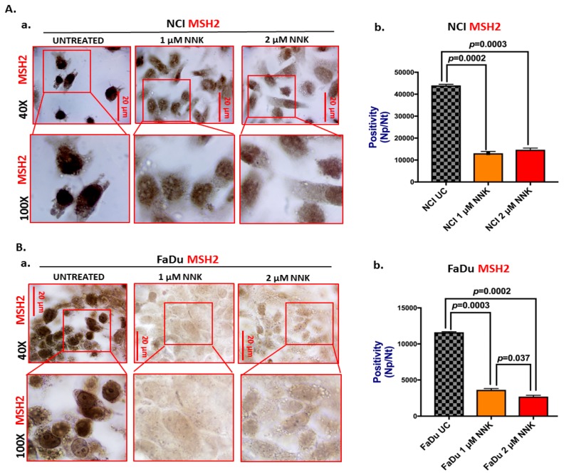
Either low or high dose of NNK reduces MSH2 expression in both lung (NCI) and head and neck (FaDu) cancer cells. Immunoperoxidase cell staining for MSH2 reveals that (A) NCI and (B) FaDU exposed to either 1 μM or 2 μM of NNK produce reduced MSH2 nuclear levels, as indicated by (A-a and B-a) the less intense MSH2 staining (scale bar: 20 μm), and (A-b and B-b) the significantly lower nuclear MSH2 levels [Np/Nt = Number of nuclear positive/total number of nuclei, means(SD)], compared to untreated controls. Data were obtained from two independent images (≥10 cells) (p values by t-test; multiple comparisons by Holm-Sidak; GraphPad Prism 7.0). Images were captured using Aperio CS2 and analyzed by Image Scope software (Leica Microsystems, Buffalo Grove, IL).

