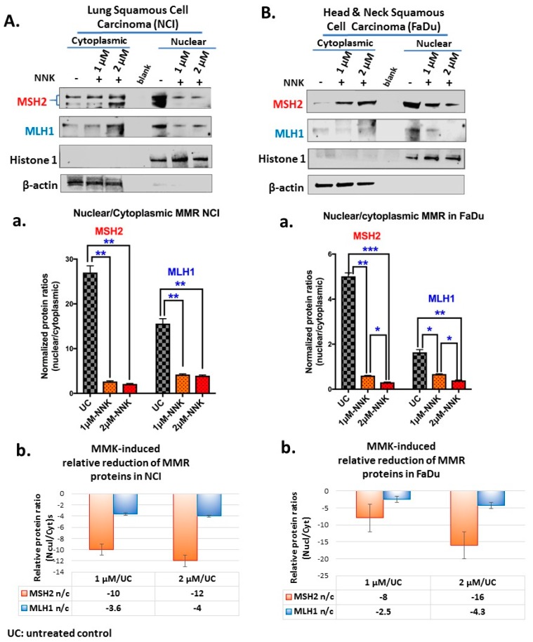
Figure 3.
Either low or high dose of NNK reduces the nuclear translocation of MMR (MSH2 and MLH1) proteins in both (A) NCI and (B) FaDu cells. Graphs depict MSH2 and MLH1 nuclear translocation ratios (nuclear/cytoplasmic protein expression levels) (A-a and B-a), in NCI and FaDu cells, respectively, exposed to 1 μM or 2 μM of NNK compared to untreated controls. (A-b and B-b) NNK-induced relative reduction of MMR nuclear translocation in NCI and FaDu cells, respectively, is demonstrated by the relative MMR n/c (nuclear/cytoplasmic) protein ratios in NNK-treated vs. untreated controls. (β-actin and Histone 1 were used to normalize cytoplasmic and nuclear protein extracts, respectively, by western blot analysis; UC: untreated controls). [Paired t-test, * p < 0.05; ** p < 0.005; *** p < 0.0005; **** p < 0.00005; GraphPad Prism 7.0; means (SD) of three independent experiments].

