Figure 4.
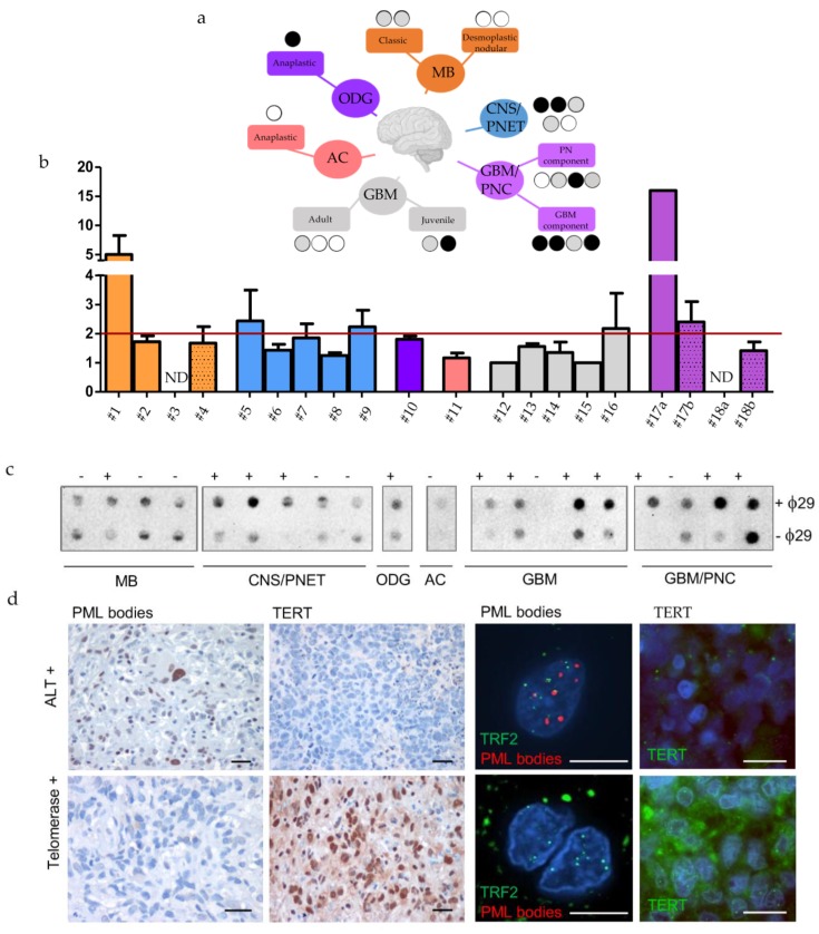
Identification of TMMs in a cohort of twenty human paediatric brain tumour. (a) Tree representation of the human cases analysed here and the TMMs identified. Black, grey and white circles indicates the results of CCA analysis (black: positive at 2 tests, grey: positive at 1 test and white: negative). (b) Identification of human brain tumors positive or negative for C-Circles, analysed by qPCR (Bars represent mean of fold changes +/- s.e.m of two independent experiments) and (c) dot blot. The red line shows the limit above which ALT is detected. MB = Medulloblastoma, CNS/PNET= Central Nervous System Primitive NeuroEctodermal Tumors, ODG = oligodendroglioma, AC = astrocytoma, GBM = glioblastoma, GBM-PNC = Glioblastoma with Primitive Neuronal Component. (d) Immunohistochemical and immunofluorescent stainings of PML bodies, TERT and PML bodies/ TRF2 in two representative cases of juvenile brain tumors, ALT or telomerase+, as indicated. Calibration bars: 10 µm.

