
Figure 2.
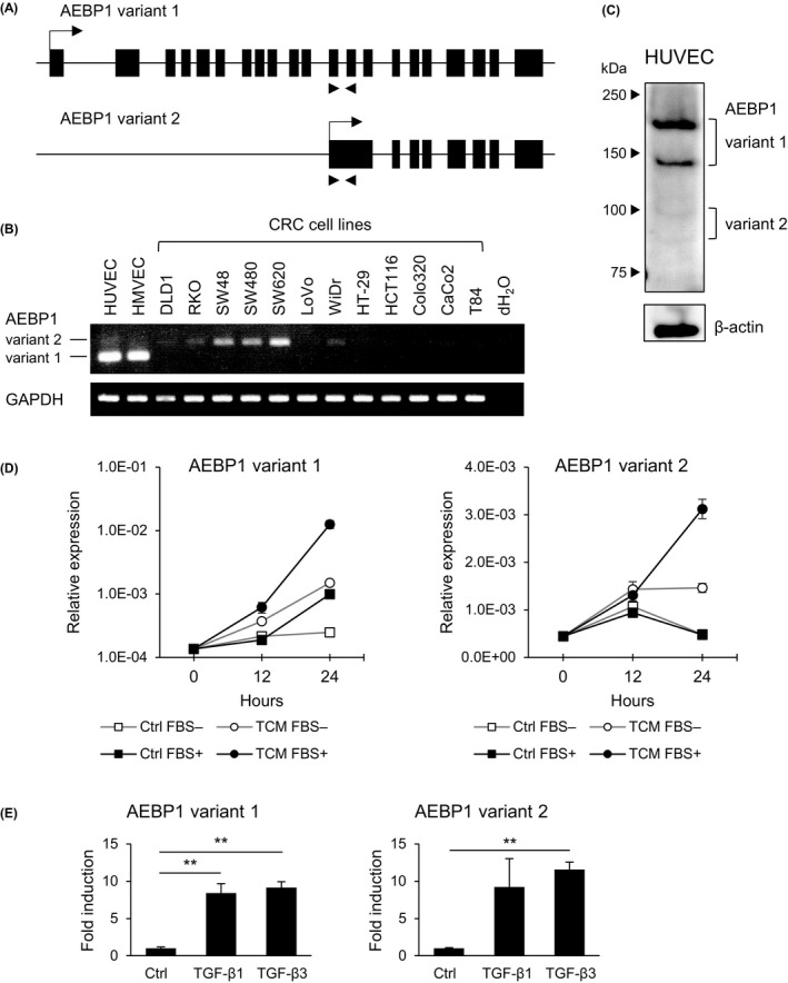
Expression of adipocyte enhancer‐binding protein 1 (AEBP1) in endothelial cells. A, Structures of genes encoding the indicated AEBP1 variants. Locations of the RT‐PCR primers used in (B) are indicated by arrows below. B, RT‐PCR of AEBP1 variants in endothelial cells and colorectal cancer (CRC) cell lines. C, Western blot analysis of AEBP1 in human umbilical vein endothelial cells (HUVECs). D, Quantitative RT‐PCR of the indicated AEBP1 variants in HUVECs treated with control medium or tumor conditioned medium (TCM) derived from DLD1 cells with or without supplemented FBS. Results are normalized to ACTB expression. Shown are means of 3 replications. E, Quantitative RT‐PCR analysis of the indicated AEBP1 variants in HUVECs treated with PBS (Ctrl), transforming growth factor (TGF)‐β1 or TGF‐β3. Shown are means of 3 replications. Error bars depict SEMs. **P < .01