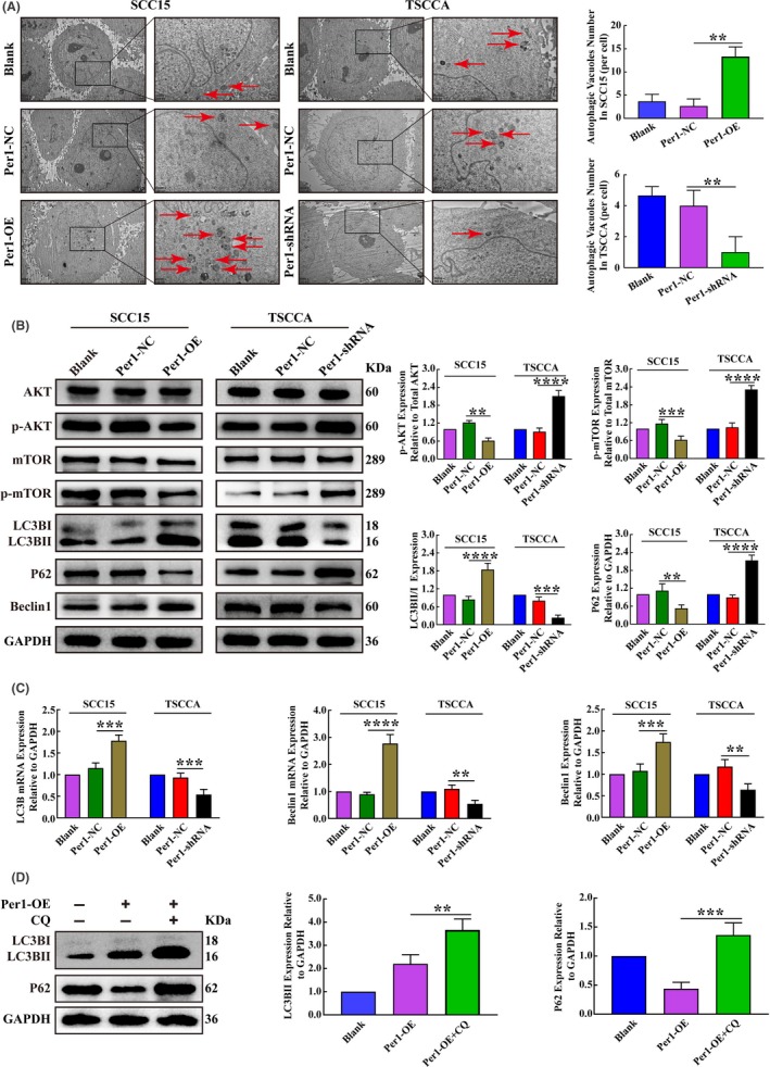
Figure 3.

Changes in autophagy and AKT/mTOR pathway activity in oral squamous cell carcinoma (OSCC) cells after the overexpression and silencing of Per1. A, Transmission electron microscopy assay showed that the autophagosome density in Per1‐OE SCC15 cells was significantly increased, while that in Per1‐shRNA TSCCA cells was significantly decreased (low magnification scale bars = 2 μm; high magnification scale bars = 1 μm). B, Western blotting showed no significant changes in the total protein levels of AKT and mTOR in Per1‐OE SCC15 cells and Per1‐shRNA TSCCA cells. In Per1‐OE SCC15 cells, the expression of p‐AKT, p‐mTOR and P62 was significantly decreased, while the LC3BII/LC3BI ratio and Beclin1 expression were significantly increased. However, in Per1‐shRNA TSCCA cells, the expression of p‐AKT, p‐mTOR and P62 was significantly increased, while the LC3BII/LC3BI ratio and Beclin1 expression were significantly decreased. C, RT‐qPCR analysis revealed that the mRNA expression of LC3B and Beclin1 was significantly increased in Per1‐OE SCC15 cells; however, the mRNA expression of LC3B and Beclin1 was significantly decreased in Per1‐shRNA TSCCA cells. D, Western blotting showed that the LC3BII and P62 expression in Per1‐OE SCC15 cells were significantly increased after addition of the lysosomal inhibitor CQ. All data represent three independent experiments. The results are shown as the mean ± SD (n ≥ 3). *P < .05; **P < .01; ***P < .001; ****P < .0001
