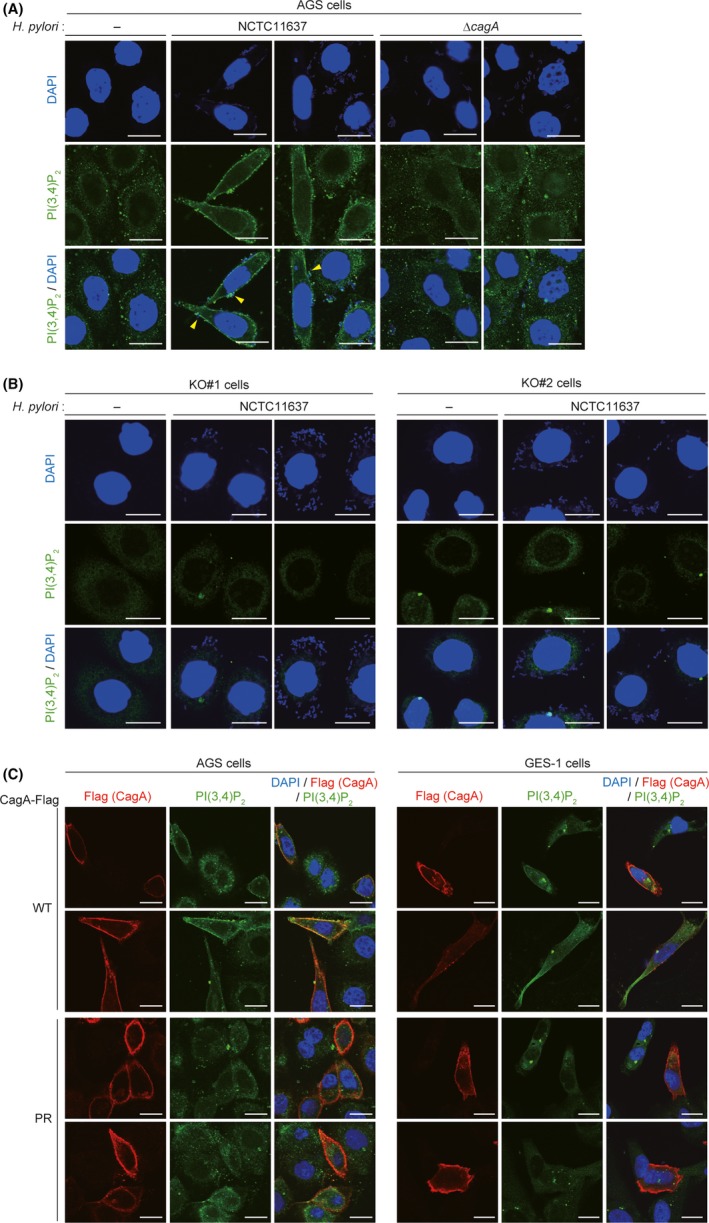
Figure 4.

Phosphatidylinositol 3,4‐diphosphate (PI(3,4)P2) accumulates in the plasma membrane of CagA‐injected cells. A, B, AGS cells (A) or AGS‐derived SHIP2‐KO KO#1 and KO#2 cells (B) were infected with the Helicobacter pylori NCTC11637 strain or its isogenic ΔcagA strain at an MOI of 100 for 6 h, and subjected to PI(3,4)P2 staining. PI(3,4)P2 was visualized in green. Cellular nuclei and H. pylori were visualized in blue by DAPI staining. Arrowheads indicate CagA‐injected cells. Scale bar, 20 μm. C, AGS or GES‐1 cells were transiently transfected with a Flag‐tagged WT‐ or PR‐CagA vector, and subjected to PI(3,4)P2 staining. PI(3,4)P2, CagA‐Flag, and cellular nuclei were visualized in green, red, and blue, respectively. Scale bar, 20 μm
