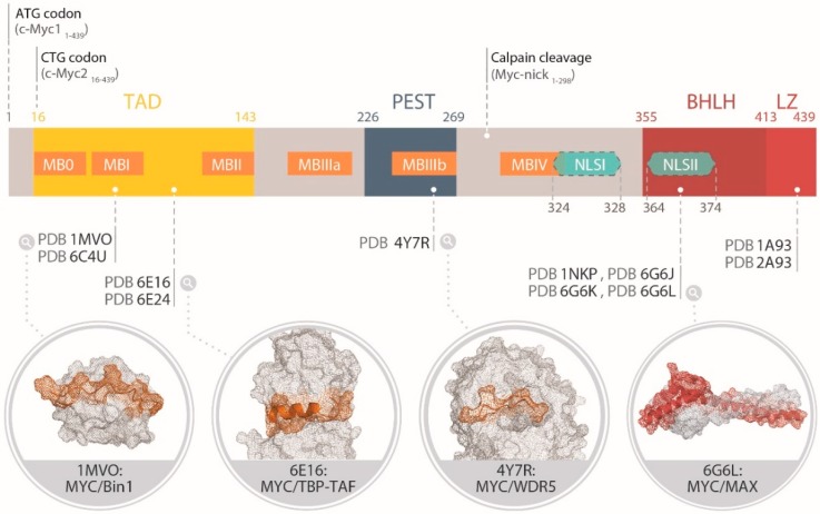
Figure 2.
Schematic representation of the modular structure of MYC (UniProtKB code P01106, https://www.uniprot.org/uniprot/P01106). The N-terminal transcription activation domain (TAD, yellow), the central region rich in proline, glutamic acid, serine and tyrosine (PEST, navy blue) and the basic region helix–loop–helix leucine zipper (LZ) domain (bHLHLZ, red) are indicated. The MYC boxes (MB 0-IV) are shown in orange and the nuclear localization sequences (NLS I-II) in cyan. The magnification bubbles show the structural models of MYC fragments (orange for MBs and red for the bHLHLZ) in complex with the interacting partners (light grey) available from the Protein Data Bank (PDB, https://www.rcsb.org) and rendered using Pymol [23]. For the TAD region: 1MVO, in complex with Bin1 [24]; 6E16 in complex with TBP-TAF [25]; 4Y7R in complex with WDR5 [26]. For the bHLHLZ, 6G6L shows the MYC/MAX heterodimer in the absence of DNA [27].

