Figure 1.
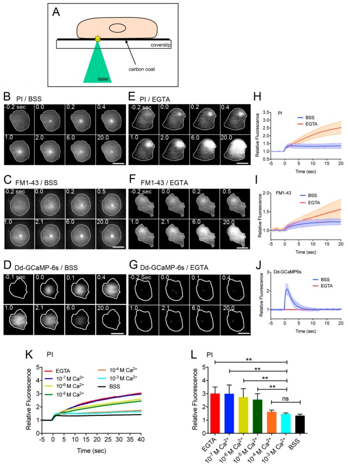
Ca2+ influx from the wound pore is essential for the wound repair. (A) To make a wound in the cell membrane, after cells were placed on a carbon-coated coverslip, a laser beam was focused on a small local spot beneath a single cell under a TIRF microscope. The wound size was set at 0.5 µm in diameter. (B,E) Typical sequences of fluorescence images of propidium iodide (PI) influx after laserporation in the presence (B) and absence (E) of Ca2+. The cells (outlined with white line) were wounded at 0 sec. (C,F) Typical sequences of fluorescence images of FM1-43 influx after laserporation in the presence and absence of Ca2+. (D,G) Typical sequences of fluorescence images of cells expressing Dd–GCaMP6s after laserporation in the presence and absence of Ca2+. (H) Time course of PI influx in the presence and absence of Ca2+. (I) Time course of FM1-43 influx after laserporation in the presence and absence of Ca2+. (J) Time course of the fluorescence intensity of cells expressing Dd–GCaMP6s after laserporation in the presence and absence of Ca2+. (K) Time courses of PI influx in the various free Ca2+concentrations in the external medium. (L) A dependency of the PI influx on the free Ca2+ concentration in the external medium. The relative fluorescence intensities 40 sec after wounding were plotted versus each Ca2+ concentration. Data are presented as mean ± SD (n = 25, each). ** p ≤ 0.0001; ns, not significant, p > 0.05. Bars, 10 µm.

