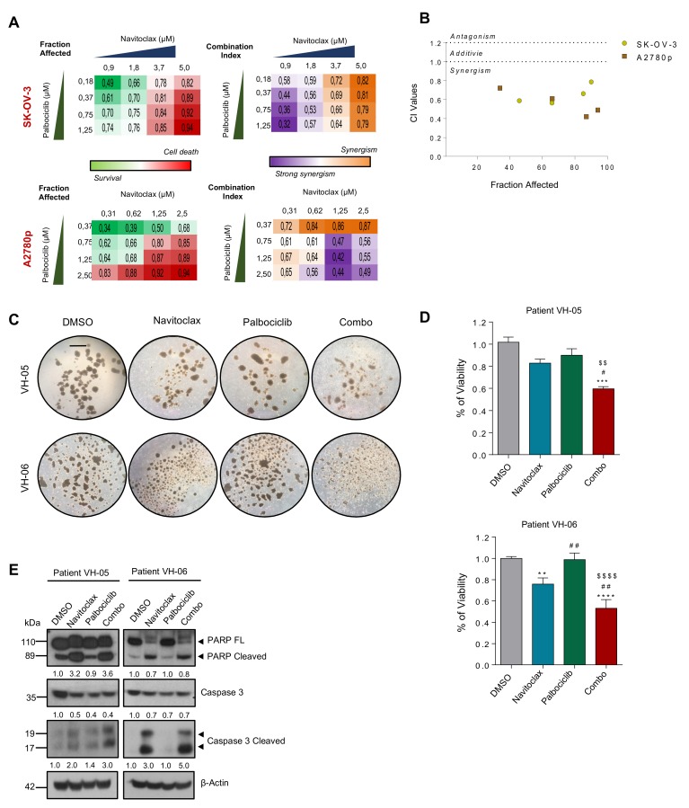
Figure 6.
BCL2 and CDK6 inhibitors cooperate to reduce the viability of patient-derived ascitic cells grown in 3D. (A) Clustering results showing fraction affected (FA) and combination index (CI) of the top combinations evaluated in SK-OV-3 and A2780p cell line using nonconstant ratio with different concentrations of Navitoclax and Palbociclib. (B) Combination index (CI) of the indicated drugs. CI was calculated by the Chou–Talay method. Data plotted are CI value of both cell lines at different FA. (C) Representative images of two patient-derived tumoral cells grown under anchorage independent conditions (tumor spheroids) and treated with Palbociclib (12 μM) and Navitoclax (4 μM) during 96 h. Bar: 100 μm (D) Viability assay (MTS) was performed on the spheres treated during 96h of the indicated drugs and the combo. Values are represented as fold change versus the control (DMSO). P-values were calculated using a one-way ANOVA analysis. * compares DMSO versus the rest of the conditions; # Navitoclax versus rest of conditions; $ Palbociclib versus Combo. # p < 0.05; **, ##, $$ p < 0.01; ***, ### p < 0.01; ****, $$$$ p < 0.001. (E) Immunoblot of the indicated protein markers to confirm the apoptosis upon simultaneous administration of the two inhibitors in primary patient-derived cells grown as spheres. β-Actin was used as a loading control.

