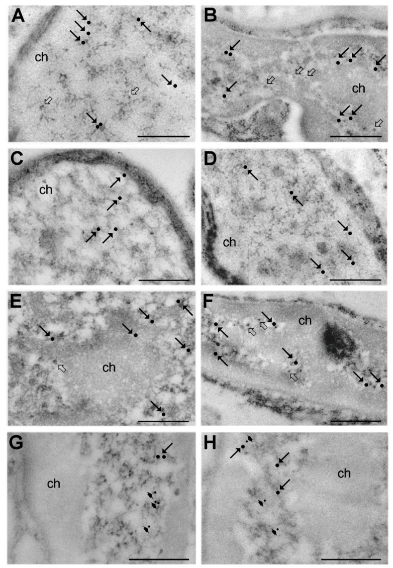
Figure 2.
Immunoelectron microscopy. SC nuclei from euthermic (A,C,E,G) and hibernating (B,D,F,H) dormice; immunolabelling for RNA polymerase II (A,B; arrows), DNA/RNA hybrid molecules (C,D; arrows), small nuclear RiboNucleoProtein ((Sm)snRNP) core protein (E,F; arrows), paired box protein 7 (Pax7) (G,H; arrows) and the myogenic differentiation transcription factor D (MyoD) (G,H; arrowheads). All antibodies specifically label perichromatin fibrils (PFs) that mostly occur at the periphery of heterochromatin clumps (ch). Perichromatin granules (PGs) are indicated by open arrows (A,B,E,F). Gold particles were digitally enhanced to improve their visibility. Bars: 500 nm.

