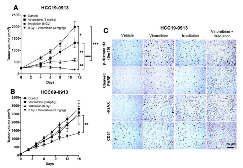
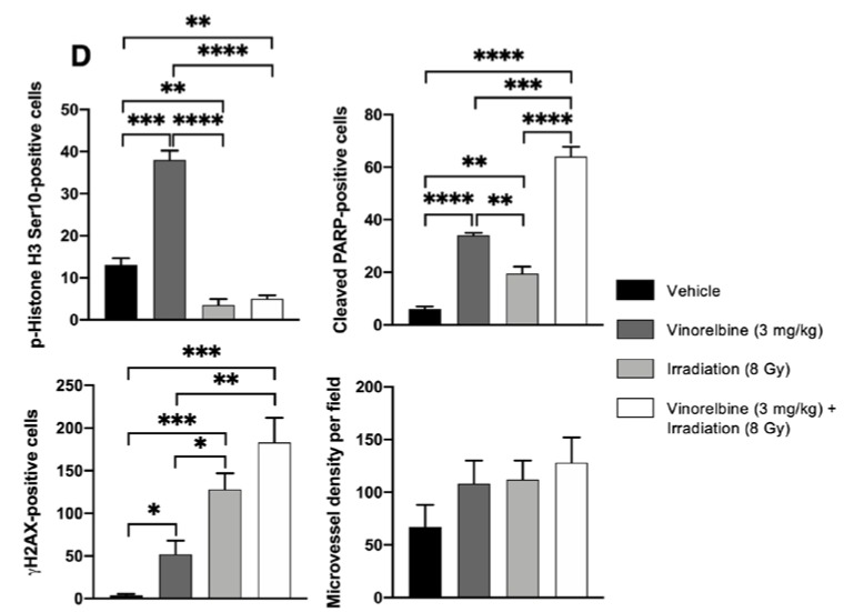
Figure 3.
Effects of localized RT, Vinorelbine, and RT/Vinorelbine on tumor growth of the HCC models. HCC19-0913 or HCC09-0913 tumors were subcutaneously implanted on both flanks and treated with vehicle, Vinorelbine, 8 Gy irradiation, or the combination of both as described in the Materials and Methods section. For irradiated mice, tumors on the right flanks were locally irradiated with 8 Gy, and the left flank tumor served as an internal control. Mean tumor volumes ± SE over time are shown (A,B). Immunohistological analysis of tumors stained with CD31, p-Histone H3 Ser10, cleaved PARP, and γH2AX as described in Appendix A (C). Representative images are shown. For p-Histone H3 Ser10, cleaved PARP, and γH2AΧ, the number of staining-positive cells among at least 500 cells per region was counted, as described in the Supplementary Materials and Methods sections, and is expressed as the number of positive cells per 1000 cells ± SE. For quantification of mean microvessel density, five random fields at a magnification of ×100 were selected for each section. The mean number of CD31-positive blood vessels per field was counted and expressed as ± SE (D). * p < 0.05; ** p < 0.01; *** p < 0.001; **** p < 0.0001, Student’s t-test.


