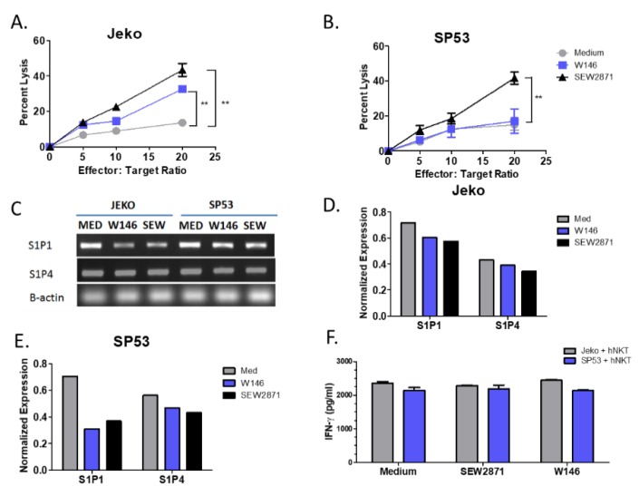
Figure 2.
Targeting of S1P1 signaling enhances NKT cell-mediated cytotoxicity of MCL. (A) Jeko and (B) SP53 cells were incubated with 10 µM SEW2871 or W146 for 72 h, washed, and co-cultured with primary NKT cells at the indicated ratios in the presence of α-GalCer (100 ng/mL) for 24 h and NKT cell mediated cell lysis was assessed by standard 51Cr-release assay. (C) MCL cell lines express S1P receptors. Expression of S1P1 and S1P4 was determined by RT-PCR after incubation with 10 µM SEW2871 (S1P1 agonist) or the S1P1 antagonist, W146, for 72 h. Expression was quantitated using densitometry for S1P1 and S1P4 in (D) Jeko and (E) SP53 relative to B-actin. (F) IFN-γ levels were determined by ELISA. Data are representative of three independent experiments. Data were analyzed by one-way ANOVA. ** p < 0.001.

